
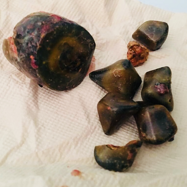
Gallstones, also called cholelithiasis, are concretions that may occur anywhere within the biliary system, most commonly within the gallbladder.
On this page:
Terminology
Gallstones (cholelithiasis) describe stone formation at any point along the biliary tree. Specific names can be given to gallstones depending on their location:
-
cholecystolithiasis: gallstones within the gallbladder
the terms cholelithiasis or gallstones have been largely used in clinical practice on their own to refer to stones in the gallbladder
choledocholithiasis: gallstones within the bile ducts
Biliary (cholesterol) microlithiasis refers to gallstones <3 mm in diameter. The term biliary microlithiasis is occasionally used as a synonym for sludge. However, this is not strictly correct as biliary sludge may include these microliths in its composition, but these are only one element of a variable mixture of crystals, proteinaceous debris, lysed cells and mucin 12.
Epidemiology
Gallstones occur in ~10% of the population, with a predominance in women (F: M = 2:1). The prevalence increases with age in both sexes 3.
Genetics may have an important role in gallstone formation. Several studies have shown an association between age-adjusted prevalence of gallstones, ethnicity 4 and family history of gallstones:
highest age-adjusted prevalence (~50%): Pima Indians, some North and South American Indians
intermediate age-adjusted prevalence (up to 20%): White (20%), and Asian population (5-20%)
lowest age-adjusted prevalence (≤5%): Africans
Risk factors
Cholesterol gallstones
female sex
middle age
positive family history
recent rapid weight loss
Also see the 5-F rule.
Pigment gallstones
-
black pigment stones
chronic hemolysis (e.g. sickle cell disease)
intestinal malabsorption (e.g. Crohn disease)
-
brown pigment stones
-
infection
bacterial
parasitic (e.g. Clonorchis sinensis)
biliary stasis
-
Clinical presentation
Gallstones may be symptomatic in only 25% of cases. The most common presentation is with biliary colic (right upper quadrant or epigastric abdominal pain or discomfort, especially after a fat-rich meal). Other symptoms include belching, bloating, flatulence, heartburn, and nausea.
Abdominal pain is often referred to the right shoulder. Patients may demonstrate this radiation to the tip of the scapula by placing their hand behind the back and thumb pointing upwards: "Collins sign". This may be useful in distinguishing gallstone pain from esophagitis, gastritis, or duodenal ulcer in ~50% of patients 5.
Pathology
There are three types of gallstones 3,4,7-10:
-
cholesterol (10%)
>50% cholesterol contents; form with supersaturation of bile, nucleation and stone growth
-
predisposing factors
diet, sedentary lifestyle, the rapid loss of weight, obesity, oral contraceptive pill, total parenteral nutrition (TPN)
ethnicity, genetic predisposition, older age, female sex
-
mixed (80%)
20-50% cholesterol content
predisposing factor: similar to cholesterol stones
-
pigment stones (10%)
<20% cholesterol content; high bilirubin content and occur when there is supersaturation of unconjugated bilirubin
-
two subtypes each with their own predisposing factors:
black pigment stones: chronic hemolysis, cirrhosis, intestinal malabsorption
brown pigment stones: infections (bacterial/parasitic) and biliary stasis
Radiographic features
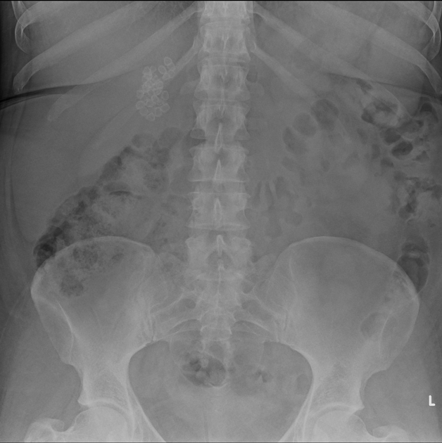
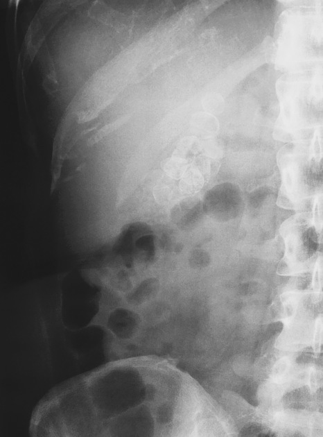
Plain radiograph
Some radiopaque gallstones may be seen on plain film:
gallstones are radiopaque only in 15-20% of cases 3
may be laminated (a.k.a. lamellated): radiopaque outline with lucent center
may have a faceted outline
may show a Mercedes-Benz sign: triradiate pattern of gas lucency
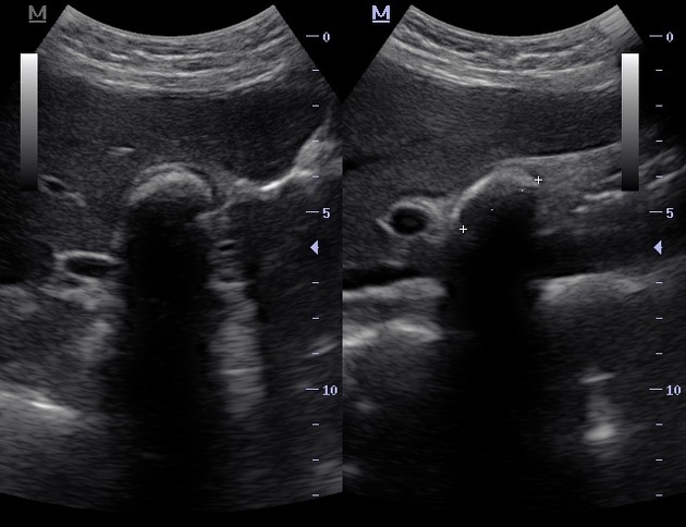
Ultrasound
Ultrasound is considered the gold standard for detecting gallstones 6:
-
greyscale ultrasound
highly reflective echogenic focus within gallbladder lumen, normally with prominent posterior acoustic shadowing regardless of pathological type (acoustic shadowing is independent of the composition and calcium content) 11
gravity-dependent movement is often seen with a change of patient position (the rolling stone sign)
-
color Doppler
may demonstrate a twinkling artifact and is particularly useful for identification of small stones
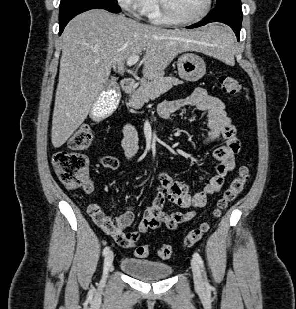
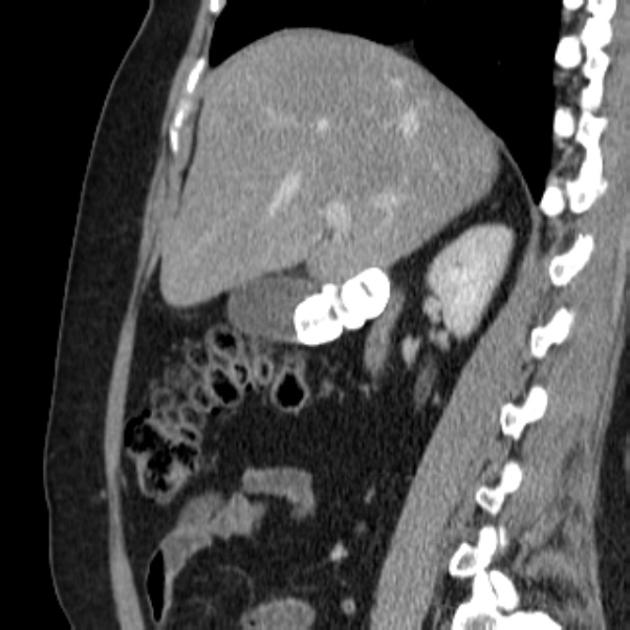
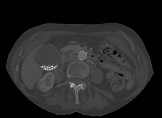
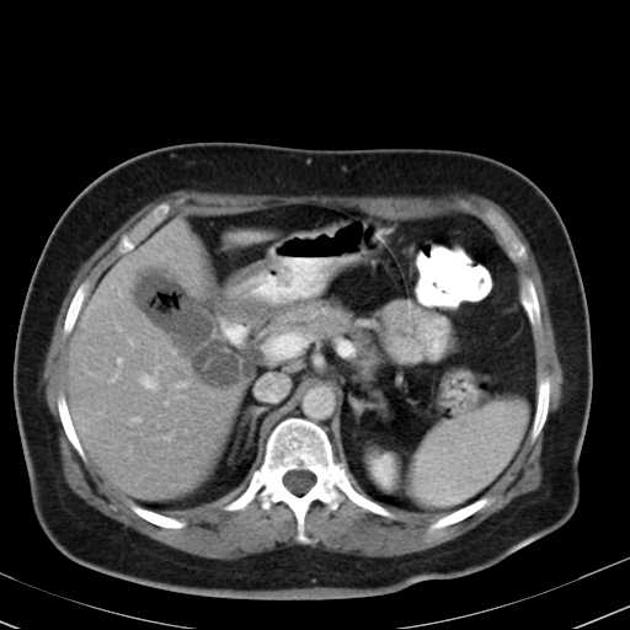
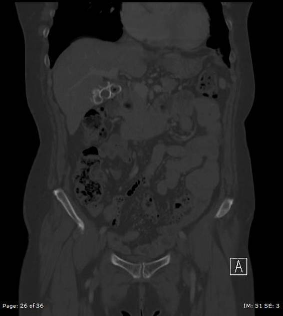
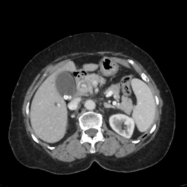
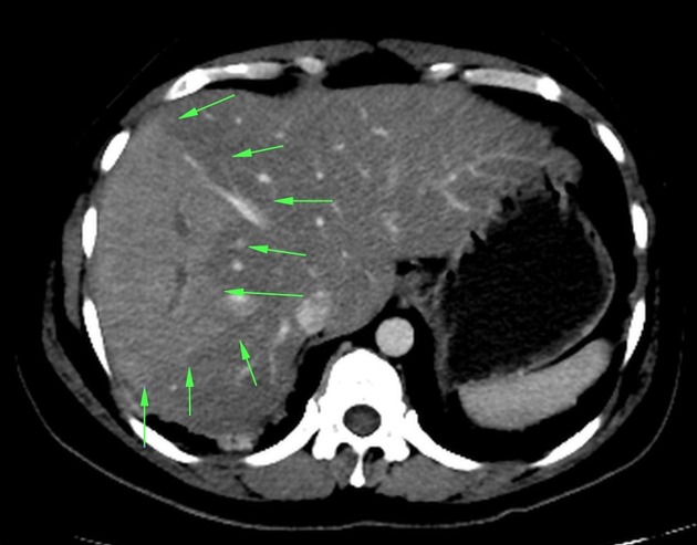
CT
Calcified gallbladder stones are hyperattenuating to bile, making them the only type to be clearly visualized on CT scan images. On CT, a high percentage of cholesterol stones are hypoattenuating relative to bile 15, and other gallstones are isodense to bile and these may not be clearly identified on CT.
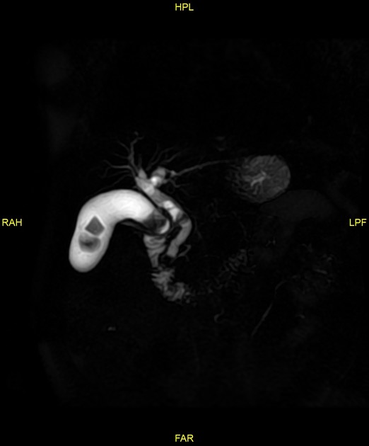
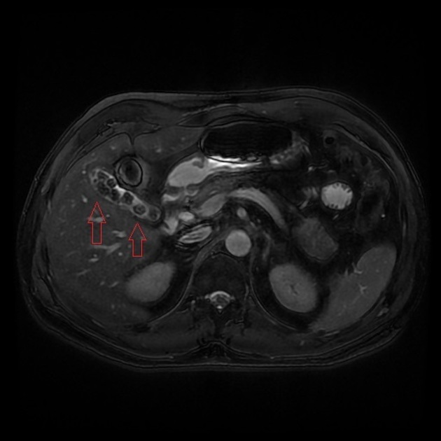
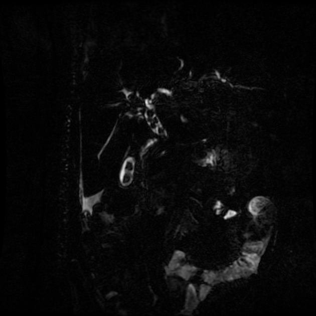
MRI
-
T1
pigmented stones (hyperintense) 14
cholesterol stones (hypointense) 14
T2: signal void or low signal outlined by markedly hyperintense bile within the gallbladder
MRCP: signal void within the gallbladder
Treatment and prognosis
Expectant management for patients with incidentally detected asymptomatic gallstones is the recommended approach. Laparoscopic cholecystectomy remains the standard treatment for gallstones. It also can be safely performed during any trimester of pregnancy when indicated 16.
Complications
biliary colic
cholecystoduodenal fistula leading to a gallstone ileus or Bouveret syndrome
increased risk of cholangiocarcinoma
Differential diagnosis
Possible imaging differential considerations in selected situations include
ingested iron tablets (pseudogallstones) on plain radiograph 13
echogenic bile (sludge) and tumefactive sludge: non-shadowing
pneumobilia on MRCP
Practical points
-
gallstone acoustic shadowing is prominent with
larger size stones (usually >3 mm for shadowing)
higher transducer frequency
focal zone at the level of gallstone
a gallbladder full of stones may paradoxically be hard to visualize (wall-echo-shadow sign)


 Unable to process the form. Check for errors and try again.