
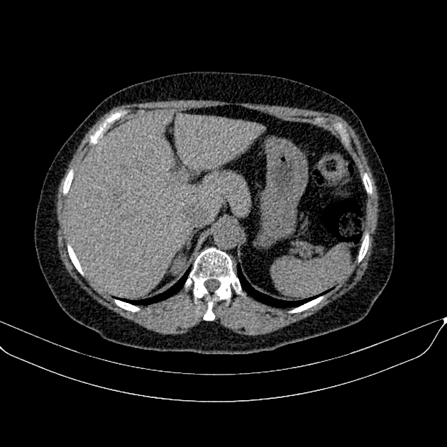
The Couinaud classification (French eponym: pronounced kwee-NO) is the most widely used system to describe functional liver anatomy. It is the preferred anatomy classification system as it divides the liver into eight independent functional units (termed segments) rather than relying on the traditional morphological description based on the external appearance of the liver.
On this page:
Terminology
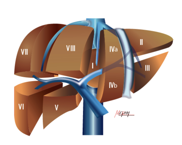
The hepatic segments were originally numbered by Roman numerals I to VIII, but the Arabic numerals 1 to 8 are now preferred 7.
Delineation of segments
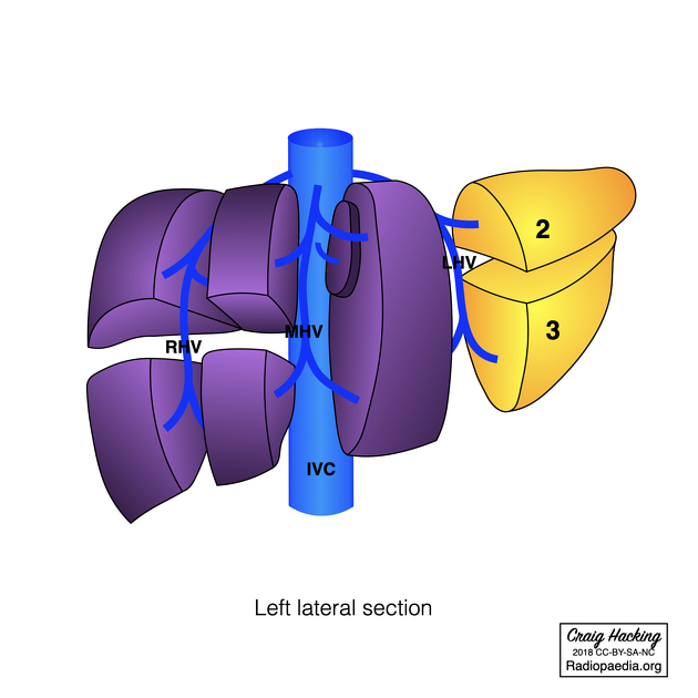
The delineation of the segments is based on the fact that each segment has its own dual vascular inflow, biliary drainage and lymphatic drainage. Generally, each segment can be conceptualised as wedge-shaped with the apex pointing towards the hepatic hilum (porta hepatis) where a single segmental branch of the portal vein, hepatic artery and bile duct enter (the portal triad). Along the boundaries of each segment there is venous outflow through the hepatic veins so that a hepatic vein drains two adjacent segments and each segment has multiple draining hepatic veins. These veins run in 3 vertical planes radiating from the intrahepatic IVC that separate 4 sections of the liver (a section is two segments on top of each other):
right hepatic vein located in the right intersegmental fissure divides the right lobe into right lateral (posterior) and right medial (anterior) sections.
middle hepatic vein lies in the main lobar fissure, divides the liver into right and left lobes (or right and left hemiliver): this vertical plane runs from the inferior vena cava to the gallbladder fossa and is known as Cantlie's line. To the right is the right medial section and to the left is the left medial section.
left hepatic vein located in the left intersegmental fissure, divides the left lobe into left medial and left lateral sections.
A horizontal plane further divides the liver, known as the portal plane where the portal vein bifurcates and becomes horizontal, dividing each section (or sector) of the liver into superior and inferior segments:
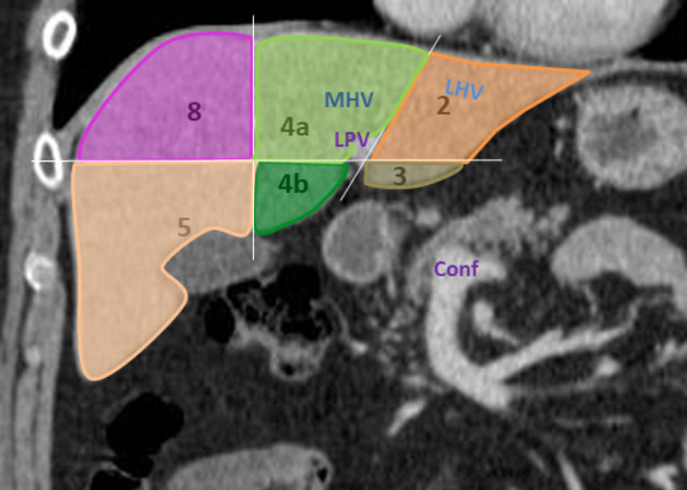
left lateral section: segment 2 above and segment 3 below the portal plane
left medial section: segment 4a above and segment 4b below the portal plane
right anterior (or medial) section: segment 8 above and segment 5 below the portal plane
right posterior (or lateral) section: segment 7 above and segment 6 below the portal plane
Segments
-
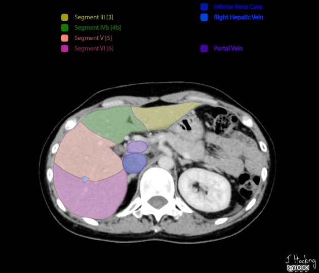
segment 1 (I) is the caudate lobe
bounded posterolaterally by the fossa for the inferior vena cava, anteriorly by the ligamentum venosum, and inferiorly by the porta hepatis
its inferior portion is subdivided into a lateral caudate process and a medial papillary process 6
may receive its supply from both the right and the left portal vein
is drained directly into the IVC by one or more small hepatic veins, explaining why it might undergo hypertrophy in certain pathologies
The remainder of the segments (2 to 8) are numbered in a clockwise fashion starting superiorly in the left hepatic lobe:
segments 2 (II) and 3 (III) are to the left of the left hepatic vein and falciform ligament with II superior and III inferior to the portal plane
-
segment 4 (IV) lies between the left and middle hepatic veins; it is subdivided into 4a (IVa) (superior) and 4b (IVb) (inferior) subsegments
easy tip: 4a is above and 4b is below the portal plane
segment 4 includes the quadrate lobe
the falciform ligament is variable in location hence is not routinely used to identify segmental boundaries
Segment 5 to 8 make up the right hepatic lobe and are easier to describe:
segment 5 (V) is located below the portal plane between the middle and right hepatic veins
segment 6 (VI) is located below the portal plane to the right of the right hepatic vein
segment 7 (VII) is located above the portal plane to the right of the right hepatic vein
segment 8 (VIII) is located above the portal plane between the middle and right hepatic veins
Each hepatic vein, therefore, drains multiple adjacent segments, those that are bounded by the hepatic vein.
A 'handy' mnemonic exists to remember the segments.
Surgical relevance
The division of the liver into self-contained units allows the surgical resection of individual segments and sections without damaging the remaining segments. Hence, for the liver to remain viable, resections occur along the hepatic veins and portal veins in the planes that define the boundaries of these segments.
Consistent universal nomenclature has been defined and promoted by the Terminology Committee of the International Hepato-Pancreato-Biliary Association (IHPBA), based on the Brisbane 2000 Terminology meeting 7:
-
first-order division anatomy
right liver or hemiliver: segments 5 - 8. Resection of these segments is termed a right hepatectomy or hemihepatectomy.
left liver or hemiliver: segments 2 - 4 (+/- segment 1). Resection of these segments is termed a left hepatectomy or hemihepatectomy (+/- segment 1).
-
second-order division anatomy
right anterior section: segments 5 and 8. Resection of these segments is termed a right anterior sectionectomy.
right posterior section: segments 6 and 7. Resection of these segments is termed a right posterior sectionectomy.
left medial section: segments 4a and 4b. Resection of these segments is termed a left medial sectionectomy or segmentectomy 4.
left lateral section: segments 2 and 3. Resection of these segments is termed a left lateral sectionectomy or bisegmentectomy 2,3.
-
third-order division anatomy
individual segments: termed segmentectomy (e.g. segmentectomy 5)
two contiguous segments: if from different sections, termed bisegmentectomy (e.g. bisegmentectomy 5, 6)
Additionally, if resection is performed of a hemiliver plus an additional adjacent section, then further nomenclature is used 7:
resection of left hemiliver plus right anterior lateral section, it is termed extended left hepatectomy or hemihepatectomy; however, the preferred term is left trisectionectomy
resection of right hemiliver plus left medial section, it is termed extended right hepatectomy or hemihepatectomy; however, the preferred term is right trisectionectomy
History and etymology
This anatomic division was first described by the French surgeon Claude Couinaud in 1957. The notion of the Couinaud liver segments being based on the arrondissements (administrative districts) of Paris is a radiological urban myth 4, but sounds cool nonetheless and is a nice way to remember the numbering.
In 2000 the Terminology Committee of the International Hepato-Pancreato-Biliary Association published a consensus hepatic nomenclature which has become rapidly adopted around the world 7,8.


 Unable to process the form. Check for errors and try again.