
Diffusion-weighted imaging
Updates to Article Attributes
Diffusion weighted-weighted imaging (DWI) is a form of MR imaging based upon measuring the random Brownian motion of water molecules within a voxel of tissue. The relationship between histology and diffusion is complex;, however, in general, highly highly cellular tissues or those with cellular swelling exhibit lower diffusion coefficients, and thus diffusion is particularly useful in tumour characterisation and cerebral ischaemia.
Terminology
A great deal of confusion exists in the way the clinicians and radiologists refer to diffusion restriction, with both groups often appearing to not actually understand what they are referring to.
The first problem is that the term "diffusion weighted-weighted imaging" is used to denote a number of different things:
- isotropic diffusion map (what most radiologists and clinicians will refer to as DWI)
-
the various pulse sequence
whichthat results in the generation ofDWIthe various images (e.g. isotropic map, b=0and, ADCmaps) - a more general term to encompass all diffusion techniques including diffusion tensor imaging
Additionally, confusion also exists in how to refer to abnormal restricted diffusion. This largely stems from the initial popularisation of DWI in stroke, which presented infarcted tissue as high signal on isotropic maps and described it merely as "restricted diffusion", implying that the rest of the brain did not demonstrate restricted diffusion, which is clearly not true. Unfortunately, this shorthand is appealing and is widespread than using the more accurate but clumsier "diffusion demonstrates greater restriction than one would expect for this tissue".
To make matters worse, many are not aware of the concept of T2 shine-through, a cause of artefactual high signal on DWI, or interpret it as a binary feature with T2 contribution to signal either present or absent when in reality there is always a T2 component even to regions with true T2 diffusion restrition.
A much safer and more accurate way of referring to diffusion restriction is to remember that we are referring to actual ADC values, and to use wording such as "the region demonstrates abnormally low ADC values (abnormal diffusion restriction)" or even "high signal on isotropic images (DWI) is confirmed by ADC maps to represent abnormal restricted diffusion".
Physics
As opposed to essentially free diffusion of water kept inside a container, diffusion of water inside brain tissue, for example, is hindered primarily by cell membrane boundaries. The overall diffusion characteristics of a single volume represent the combined water diffusion in a number of compartments:
- diffusion within the intracellular fluid
- within the cytoplasm generally
- within organelles
- diffusion within extracellular fluid
- interstitial fluid
- intravascular
- lymphatic
- various biological cavities, e.g. ventricles of the brain
- diffusion between intra- and extracellular compartments
The contribution of each one of these will depend on the tissue and pathology. For example, in acute cerebral infarction it is believed that the decrease in ADC values is the result of a combination of water moving into the intracellular compartment (where its diffusion is more impeded by organelles than it is in the extracellular space) and the resulting cellular swelling narrowing the the extracellular space 6. Similar mechanisms result in low ADC values in highly cellular tumours (e.g. small round blue cell tumours (e.g. lymphoma / PNET) and high grade gliomas (GBM)).
The further an individual water molecule diffuses during the sequence the more it will be exposed to varying gradient strength and the more it will be dephased reducing the amount of signal returned. This occurs at a much smaller scale than a single voxel. The strength of this effect (in other words how much the signal will be attenuated by diffusion) is determined by the B value.
Clinical application
DWDiffusion-weighted imaging has a major role in the following clinical situations 3-5:
- early identification of ischemic stroke
- differentiation of acute from chronic stroke
- differentiation of acute stroke from other stroke mimics
- differentiation of epidermoid cyst from an arachnoid cyst
- differentiation of abscess from necrotic tumors
- assessment of cortical lesions in
CJDCreutzfeld-Jacob disease (CJD) - differentiation of herpes encephalitis from diffuse temporal gliomas
- assessment of the extent of diffuse axonal injury
- grading of diffuse gliomas and meningiomas
(need further study) - assessment of active demyelination
MRI sequence
Figure 1 depictsA variety of techniques for generating diffusion maps have been developed. By far the most commonly used technique relies on a spin echo-echo echoplanar sequence with(SE-EPI) although non-EPI techniques (e.g. turbo spine echo) are also available and are of use particularly where tissue is adjacent or within bone where T2* effects cause artefact, distortion and signal loss on EPI sequences 7,8.
General principle of diffusion gradients added-weighted imaging
The fundamental idea behind diffusion-weighted imaging is the attenuation of T2* signal based on how easily water molecules are able to diffuse in that region. The gradient coil usedmore easily water can diffuse (i.e. the further a water molecule can move around during the sequence) the less initial T2* signal will remain. For example, water within CSF can diffuse very easily and therefore very little signal remains and the ventricles appear black. In contrast, water within brain parenchyma cannot move as easily due to producecell membranes getting in the way and therefore the initial T2* signal of the brain is only somewhat attenuated. An important consequence of this is that if a region of the brain has zero T2* signal it cannot, regardless of the diffusion need not becharacteristics of that tissue, show signal on isotropic diffusion-weighted images.
The way in which diffusion information is extracted from the tissue is to first obtain a separateT2* weighted image with no diffusion attenuation. This is known as the b=0 image.
Next, the ease with which water can diffuse is assessed in various directions; the minimum is 3 orthogonal directions (X, Y and Z) and we will use this for the rest of this explanation.
This is done by applying a strong gradient or gradients from those used for spatial encodingsymmetrically on either side of the 180-degree pulse. The The degree of diffusion weighting is dependent primarily on the area under the diffusion gradients (amplitude and duration) and on the interval between the gradients. Other factors include
Stationary water molecules acquire phase information by the effectapplication of the spatial localization gradients andfirst gradient. After the size180-degree pulse, however, they are exposed to the exact same gradient (because they have not changed location) which undoes all the effects of the voxelsfirst (since they have flipped 180-degrees).
-
stationary water molecule - unaffected byHence at thediffusion gradients and hence retaintime the echo is generated they have retained their signal. -
movingMoving
watermolecule - acquiremolecules, on the other hand, acquire phase information by the first gradient but as they are moving when they are exposed to the second gradient they are not in the same location and thus are not exposed to precisely the same gradient after the 180-degree pulse. Hence they are not rephasedby the second, hence losingand they lose some of their signal.
Generating isotropic DWI and ADC maps
The aforementioned process generates four sets images: a T2* b=0 image and three diffuse weighted images (one for each of X, Y and Z direction) with the T2* signal attenuated according to how easily water can diffuse in that direction.
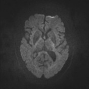
These images can then be combined arithmetically to generate maps that are devoid of directional information (isotropic) generating isotripc diffusion-weighted images (what we usually simply refer to as DWI) and apparent diffusion coefficient (ADC) maps.
To generate the isotropic DWI maps the geometric mean of the direction-specific images is calculated.
The ADC map, in contrast, is related to the natural logarithm (ln) of the isotropic DWI divided by the initial T2* signal (b=0).
See also
-<p><strong>Diffusion weighted imaging (DWI)</strong> is a form of MR imaging based upon measuring the random Brownian motion of water molecules within a voxel of tissue. The relationship between histology and diffusion is complex; however, in general, highly cellular tissues or those with cellular swelling exhibit lower diffusion coefficients, and thus diffusion is particularly useful in tumour characterisation and cerebral ischaemia. </p><h4>Terminology</h4><p>A great deal of confusion exists in the way the clinicians and radiologists refer to diffusion restriction, with both groups often appearing to not actually understand what they are referring to.</p><p>The first problem is that the term "diffusion weighted imaging" is used to denote a number of different things: </p><ol>-<li>isotropic diffusion map (what most radiologists will refer to as DWI)</li>-<li>sequence which results in generation of DWI, b=0 and ADC maps</li>- +<p><strong>Diffusion-weighted imaging (DWI)</strong> is a form of MR imaging based upon measuring the random Brownian motion of water molecules within a voxel of tissue. The relationship between histology and diffusion is complex, however, in general highly cellular tissues or those with cellular swelling exhibit lower diffusion coefficients, and thus diffusion is particularly useful in tumour characterisation and cerebral ischaemia. </p><h4>Terminology</h4><p>A great deal of confusion exists in the way the clinicians and radiologists refer to diffusion restriction, with both groups often appearing to not actually understand what they are referring to.</p><p>The first problem is that the term "diffusion-weighted imaging" is used to denote a number of different things: </p><ol>
- +<li>isotropic diffusion map (what most radiologists and clinicians will refer to as DWI)</li>
- +<li>the various pulse sequence that results in the generation of the various images (e.g. isotropic map, b=0, ADC)</li>
-</ol><p>Additionally, confusion also exists in how to refer to abnormal restricted diffusion. This largely stems from the initial popularisation of DWI in stroke, which presented infarcted tissue as high signal on isotropic maps and described it merely as "restricted diffusion", implying that the rest of the brain did not demonstrate restricted diffusion, which is clearly not true. Unfortunately, this shorthand is appealing and is widespread than using the more accurate but clumsier "diffusion demonstrates greater restriction than one would expect for this tissue".</p><p>To make matters worse, many are not aware of the concept of <a href="/articles/t2-shine-through">T2 shine-through</a>, a cause of artefactual high signal on DWI.</p><p>A much safer and more accurate way of referring to diffusion restriction is to remember that we are referring to actual ADC values, and to use wording such as "the region demonstrates abnormally low ADC values (abnormal diffusion restriction)" or even "high signal on isotropic images (DWI) is confirmed by ADC maps to represent abnormal restricted diffusion".</p><h4>Physics</h4><p>As opposed to essentially free diffusion of water kept inside a container, diffusion of water inside brain tissue, for example, is hindered primarily by cell membrane boundaries. The overall diffusion characteristics of a single volume represent the combined water diffusion in a number of compartments: </p><ul>- +</ol><p>Additionally, confusion also exists in how to refer to abnormal restricted diffusion. This largely stems from the initial popularisation of DWI in stroke, which presented infarcted tissue as high signal on isotropic maps and described it merely as "restricted diffusion", implying that the rest of the brain did not demonstrate restricted diffusion, which is clearly not true. Unfortunately, this shorthand is appealing and is widespread than using the more accurate but clumsier "diffusion demonstrates greater restriction than one would expect for this tissue".</p><p>To make matters worse, many are not aware of the concept of <a href="/articles/t2-shine-through">T2 shine-through</a>, a cause of artefactual high signal on DWI, or interpret it as a binary feature with T2 contribution to signal either present or absent when in reality there is always a T2 component even to regions with true T2 diffusion restrition. </p><p>A much safer and more accurate way of referring to diffusion restriction is to remember that we are referring to actual ADC values, and to use wording such as "the region demonstrates abnormally low ADC values (abnormal diffusion restriction)" or even "high signal on isotropic images (DWI) is confirmed by ADC maps to represent abnormal restricted diffusion".</p><h4>Physics</h4><p>As opposed to essentially free diffusion of water kept inside a container, diffusion of water inside brain tissue, for example, is hindered primarily by cell membrane boundaries. The overall diffusion characteristics of a single volume represent the combined water diffusion in a number of compartments: </p><ul>
-</ul><p>The contribution of each one of these will depend on the tissue and pathology. For example, in acute cerebral infarction it is believed that the decrease in ADC values is the result of a combination of water moving into the intracellular compartment (where its diffusion is more impeded by organelles than it is in the extracellular space) and the resulting cellular swelling narrowing the the extracellular space <sup>6</sup>. Similar mechanisms result in low ADC values in highly cellular tumours (e.g. <a href="/articles/small-round-blue-cell-tumours">small round blue cell tumours</a> (e.g. lymphoma / PNET) and high grade gliomas (<a href="/articles/glioblastoma">GBM</a>)). </p><p>The further an individual water molecule diffuses during the sequence the more it will be exposed to varying gradient strength and the more it will be dephased reducing the amount of signal returned. This occurs at a much smaller scale than a single voxel. The strength of this effect (in other words how much the signal will be attenuated by diffusion) is determined by the B value. </p><h4>Clinical application</h4><p>DW imaging has a major role in the following clinical situations <sup>3-5</sup>: </p><ul>- +</ul><p>The contribution of each one of these will depend on the tissue and pathology. For example, in acute cerebral infarction it is believed that the decrease in ADC values is the result of a combination of water moving into the intracellular compartment (where its diffusion is more impeded by organelles than it is in the extracellular space) and the resulting cellular swelling narrowing the the extracellular space <sup>6</sup>. Similar mechanisms result in low ADC values in highly cellular tumours (e.g. <a href="/articles/small-round-blue-cell-tumours">small round blue cell tumours</a> (e.g. lymphoma / PNET) and high grade gliomas (<a href="/articles/glioblastoma">GBM</a>)). </p><p>The further an individual water molecule diffuses during the sequence the more it will be exposed to varying gradient strength and the more it will be dephased reducing the amount of signal returned. This occurs at a much smaller scale than a single voxel. The strength of this effect (in other words how much the signal will be attenuated by diffusion) is determined by the B value. </p><h4>Clinical application</h4><p>Diffusion-weighted imaging has a major role in the following clinical situations <sup>3-5</sup>: </p><ul>
-<li>differentiation of <a href="/articles/intracranial-epidermoid-cyst">epidermoid cyst</a> from <a href="/articles/arachnoid-cyst">arachnoid cyst</a>- +<li>differentiation of <a href="/articles/intracranial-epidermoid-cyst">epidermoid cyst</a> from an <a href="/articles/arachnoid-cyst">arachnoid cyst</a>
-<li>assessment of cortical lesions in <a href="/articles/creutzfeldt-jakob-disease">CJD</a>-</li>- +<li>assessment of cortical lesions in <a href="/articles/creutzfeldt-jakob-disease">Creutzfeld-Jacob disease (CJD</a>)</li>
-<li>grading of gliomas and meningiomas (need further study)</li>- +<li>grading of <a title="Diffuse glioma" href="/articles/diffuse-glioma">diffuse gliomas</a> and <a title="Meningiomas" href="/articles/meningioma">meningiomas</a>
- +</li>
-</ul><ol></ol><h4>MRI sequence</h4><p>Figure 1 depicts a spin echo sequence with diffusion gradients added. The gradient coil used to produce the diffusion need not be a separate gradient or gradients from those used for spatial encoding. The degree of diffusion weighting is dependent primarily on the area under the diffusion gradients and on the interval between the gradients. Other factors include the effect of the spatial localization gradients and the size of the voxels. </p><ul>-<li>stationary water molecule - unaffected by the diffusion gradients and hence retain their signal.</li>-<li>moving water molecule - acquire phase information by the first gradient but are not rephased by the second, hence losing their signal.</li>-</ul><h4>See also</h4><ul>- +</ul><ol></ol><h4>MRI sequence</h4><p>A variety of techniques for generating diffusion maps have been developed. By far the most commonly used technique relies on a spin-echo echoplanar sequence (SE-EPI) although non-EPI techniques (e.g. turbo spine echo) are also available and are of use particularly where tissue is adjacent or within bone where T2* effects cause artefact, distortion and signal loss on EPI sequences <sup>7,8</sup>. </p><h5>General principle of diffusion-weighted imaging</h5><p>The fundamental idea behind diffusion-weighted imaging is the attenuation of T2* signal based on how easily water molecules are able to diffuse in that region. The more easily water can diffuse (i.e. the further a water molecule can move around during the sequence) the less initial T2* signal will remain. For example, water within CSF can diffuse very easily and therefore very little signal remains and the ventricles appear black. In contrast, water within brain parenchyma cannot move as easily due to cell membranes getting in the way and therefore the initial T2* signal of the brain is only somewhat attenuated. An important consequence of this is that if a region of the brain has zero T2* signal it cannot, regardless of the diffusion characteristics of that tissue, show signal on isotropic diffusion-weighted images. </p><p>The way in which diffusion information is extracted from the tissue is to first obtain a T2* weighted image with no diffusion attenuation. This is known as the b=0 image. </p><p>Next, the ease with which water can diffuse is assessed in various directions; the minimum is 3 orthogonal directions (X, Y and Z) and we will use this for the rest of this explanation.</p><p>This is done by applying a strong gradient symmetrically on either side of the 180-degree pulse. The degree of diffusion weighting is dependent primarily on the area under the diffusion gradients (amplitude and duration) and on the interval between the gradients. </p><p>Stationary water molecules acquire phase information by the application of the first gradient. After the 180-degree pulse, however, they are exposed to the exact same gradient (because they have not changed location) which undoes all the effects of the first (since they have flipped 180-degrees). Hence at the time the echo is generated they have retained their signal.</p><p>Moving water molecules, on the other hand, acquire phase information by the first gradient but as they are moving when they are exposed to the second gradient they are not in the same location and thus are not exposed to precisely the same gradient after the 180-degree pulse. Hence they are not rephased and they lose some of their signal. The further they are able to move the less successfully they will be rephased and the less signal will remain. </p><h5>Generating isotropic DWI and ADC maps</h5><p>The aforementioned process generates four sets images: a T2* b=0 image and three diffuse weighted images (one for each of X, Y and Z direction) with the T2* signal attenuated according to how easily water can diffuse in that direction. </p><p>These images can then be combined arithmetically to generate maps that are devoid of directional information (isotropic) generating isotripc diffusion-weighted images (what we usually simply refer to as DWI) and apparent diffusion coefficient (ADC) maps. </p><p>To generate the isotropic DWI maps the geometric mean of the direction-specific images is calculated. </p><p>The ADC map, in contrast, is related to the natural logarithm (ln) of the isotropic DWI divided by the initial T2* signal (b=0). </p><p></p><h4>See also</h4><ul>
References changed:
- 7. Harris AD, Pereira RS, Mitchell JR, Hill MD, Sevick RJ, Frayne R. A comparison of images generated from diffusion-weighted and diffusion-tensor imaging data in hyper-acute stroke. (2004) Journal of magnetic resonance imaging : JMRI. 20 (2): 193-200. <a href="https://doi.org/10.1002/jmri.20116">doi:10.1002/jmri.20116</a> - <a href="https://www.ncbi.nlm.nih.gov/pubmed/15269943">Pubmed</a> <span class="ref_v4"></span>
- 8. B. De Foer, J.-P. Vercruysse, B. Pilet, J. Michiels, R. Vertriest, M. Pouillon, T. Somers, J.W. Casselman, E. Offeciers. Single-Shot, Turbo Spin-Echo, Diffusion-Weighted Imaging versus Spin-Echo-Planar, Diffusion-Weighted Imaging in the Detection of Acquired Middle Ear Cholesteatoma. (2006) American Journal of Neuroradiology. 27 (7): 1480. <a href="https://www.ncbi.nlm.nih.gov/pubmed/16908562">Pubmed</a> <span class="ref_v4"></span>
Image 6 MRI (@ 6 B values) ( update )

Image 7 MRI (@ 6 B values) ( update )

Image 8 MRI (@ 6 B values) ( update )
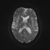
Image 9 MRI (@ 6 B values) ( update )

Image 10 MRI (@ 6 B values) ( update )

Image 11 MRI (@ 6 B values) ( update )


 Unable to process the form. Check for errors and try again.