
Gartner duct cysts develop from embryologic remnants of the Wolffian (mesonephric) duct. They are often noticed incidentally on ultrasound or MRI.
On this page:
Epidemiology
Associations
Gartner duct cysts most often are isolated findings, but can also be associated with abnormalities of the metanephric urinary system or in Herlyn-Werner-Wunderlich syndrome 4-7
ipsilateral renal dysplasia 6
Clinical presentation
They may cause mass effect on adjacent structures.
Pathology
Location
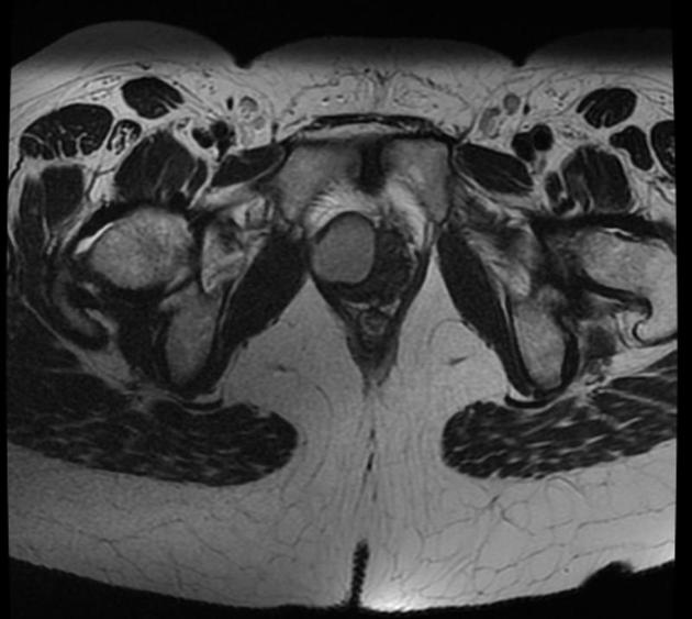
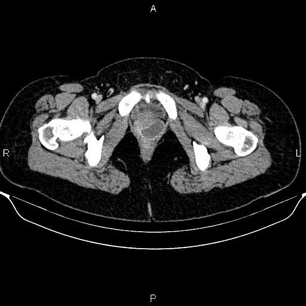
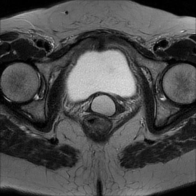
Gartner duct cysts are located in the anterolateral wall of the proximal (superior) portion of the vagina 2 and are typically above the level of the most inferior aspect of the pubic symphysis.
Microscopic appearance
Like other cysts, they are lined with non-mucinous cuboidal or columnar epithelium.
Radiographic features
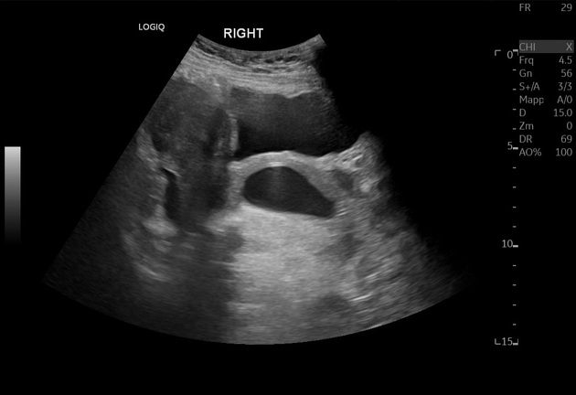
Typically, they are simple cystic lesions arising from the anterolateral aspect of the superior vagina. They are usually small (<2 cm) although occasionally they can become very large (up to several centimeters) 3. Signal characteristics can vary depending on the presence of hemorrhage.
Treatment and prognosis
Complications
In rare cases with larger cysts, dyspareunia and problems in obstetric delivery have been described 3.
Only one case of malignant transformation of Gartner's duct cyst is described 9.
Differential diagnosis
General imaging differential considerations include:
Bartholin gland cyst: their location at or below the level of the pubic symphysis and usually arising from the posterolateral wall of the vagina; this helps to differentiate them from Gartner duct cysts
urethral diverticulum: located around the urethra


 Unable to process the form. Check for errors and try again.