
Maisonneuve fracture
Updates to Article Attributes
Maisonneuve fracture refers to a combination of a fracture of the proximal fibula together with an unstable ankle injury (widening of the ankle mortise on x-ray), often comprising ligamentous injury (distal tibiofibular syndesmosis, deltoid ligament) and/or fracture of the medial malleolus. It is caused by a pronation-external rotation mechanism.
Radiographic features
Plain radiograph
Ankle views may show a fracture of the medial malleolus or widening of the medial ankle joint space due to deltoid ligament injury, as well as widening of the distal tibiofibular syndesmosis. When these ankle injury types are seen without a fracture of the lateral malleolus, further imaging of the entire fibula is recommended.
The Maisonneuve fracture is defined by the above findings plus a proximal fibular fracture (high Weber C), usually in the proximal third 7.
Treatment and prognosis
Although management is variable depending on the complexity of injuries, this type of fracture pattern is generally managed by operative treatment. Specific aims generally include:
- internal fixation of the distal tibiofibular syndesmosis 6
-
commonly achieved by trans-syndesmotic screws. Alternative
stabilizationstabilisation mechanisms exist (e.g. bioabsorbable constructs, syndesmotic staples, hooks, or cerclage wires) - in some cases, internal fixation of a posterior malleolar fracture fragment may result in sufficient
stabilizationstabilisation - fixation screws may or may not be removed after several weeks of healing 6
-
commonly achieved by trans-syndesmotic screws. Alternative
- reduction and
stabilizationstabilisation of medial malleolus fracture and/or ligamentous injuries 6- ligamentous injuries may be managed non-operatively
-
reduction and
stabilizationstabilisation of the fibular fracture- fracture involving distal 2/3 of the fibula may compromise ankle mortise, and so may benefit from surgery 6
- fracture involving proximal 1/3 fibula often managed non-operatively
History and etymology
It is named after Jules Germain Francois Maisonneuve, French surgeon (1809-1897) 1,4.
-<p><strong>Maisonneuve fracture</strong> refers to a combination of a fracture of the proximal <a href="/articles/fibula">fibula</a> together with an unstable ankle injury (widening of the ankle mortise on x-ray), often comprising ligamentous injury (distal <a href="/articles/distal-tibiofibular-syndesmosis">tibiofibular syndesmosis</a>, <a href="/articles/deltoid-ligament-of-the-ankle-1">deltoid ligament</a>) and/or fracture of the medial malleolus. It is caused by a pronation-external rotation <a href="/articles/lauge-hansen-classification-of-ankle-injury">mechanism</a>.</p><h4>Radiographic features</h4><h5>Plain radiograph</h5><p>Ankle views may show a fracture of the medial malleolus or widening of the medial ankle joint space due to deltoid ligament injury, as well as widening of the distal <a href="/articles/distal-tibiofibular-syndesmosis">tibiofibular syndesmosis</a>. When these ankle injury types are seen without a fracture of the lateral malleolus, further imaging of the entire fibula is recommended.</p><p>The Maisonneuve fracture is defined by the above findings plus a proximal fibular fracture (high <a href="/articles/weber-classification-of-ankle-fractures">Weber C</a>), usually in the proximal third <sup>7</sup>.</p><h4>Treatment and prognosis</h4><p>Although management is variable depending on the complexity of injuries, this type of fracture pattern is generally managed by operative treatment. Specific aims generally include:</p><ol>-<li>internal fixation of the distal <a href="/articles/distal-tibiofibular-syndesmosis">tibiofibular syndesmosis</a> <sup>6</sup><ul>-<li>-<a href="/articles/distal-tibiofibular-syndesmosis"></a>commonly achieved by trans-syndesmotic screws. Alternative stabilization mechanisms exist (e.g. bioabsorbable constructs, syndesmotic staples, hooks, or cerclage wires)</li>-<li>in some cases, internal fixation of a posterior malleolar fracture fragment may result in sufficient stabilization</li>-<li>fixation screws may or may not be removed after several weeks of healing <sup>6</sup>-</li>-</ul>-</li>-<li>reduction and stabilization of medial malleolus fracture and/or ligamentous injuries <sup>6</sup><ul><li>-<sup></sup>ligamentous injuries may be managed non-operatively</li></ul>-</li>-<li>-<sup></sup>reduction and stabilization of the fibular fracture<ul>-<li>fracture involving distal 2/3 of the fibula may compromise ankle mortise, and so may benefit from surgery <sup>6</sup>-</li>-<li>fracture involving proximal 1/3 fibula often managed non-operatively</li>-</ul>-</li>- +<p><strong>Maisonneuve fracture</strong> refers to a combination of a fracture of the proximal <a href="/articles/fibula">fibula</a> together with an unstable ankle injury (widening of the ankle mortise on x-ray), often comprising ligamentous injury (distal <a href="/articles/distal-tibiofibular-syndesmosis">tibiofibular syndesmosis</a>, <a href="/articles/deltoid-ligament-of-the-ankle-1">deltoid ligament</a>) and/or fracture of the medial malleolus. It is caused by a pronation-external rotation <a href="/articles/lauge-hansen-classification-of-ankle-injury">mechanism</a>.</p><h4>Radiographic features</h4><h5>Plain radiograph</h5><p>Ankle views may show a fracture of the medial malleolus or widening of the medial ankle joint space due to deltoid ligament injury, as well as widening of the distal <a href="/articles/distal-tibiofibular-syndesmosis">tibiofibular syndesmosis</a>. When these ankle injury types are seen without a fracture of the lateral malleolus, further imaging of the entire fibula is recommended.</p><p>The Maisonneuve fracture is defined by the above findings plus a proximal fibular fracture (high <a href="/articles/weber-classification-of-ankle-fractures">Weber C</a>), usually in the proximal third <sup>7</sup>.</p><h4>Treatment and prognosis</h4><p>Although management is variable depending on the complexity of injuries, this type of fracture pattern is generally managed by operative treatment. Specific aims generally include:</p><ol>
- +<li>internal fixation of the distal <a href="/articles/distal-tibiofibular-syndesmosis">tibiofibular syndesmosis</a> <sup>6</sup><ul>
- +<li>
- +<a href="/articles/distal-tibiofibular-syndesmosis"></a>commonly achieved by trans-syndesmotic screws. Alternative stabilisation mechanisms exist (e.g. bioabsorbable constructs, syndesmotic staples, hooks, or cerclage wires)</li>
- +<li>in some cases, internal fixation of a posterior malleolar fracture fragment may result in sufficient stabilisation</li>
- +<li>fixation screws may or may not be removed after several weeks of healing <sup>6</sup>
- +</li>
- +</ul>
- +</li>
- +<li>reduction and stabilisation of medial malleolus fracture and/or ligamentous injuries <sup>6</sup><ul><li>
- +<sup></sup>ligamentous injuries may be managed non-operatively</li></ul>
- +</li>
- +<li>
- +<sup></sup>reduction and stabilisation of the fibular fracture<ul>
- +<li>fracture involving distal 2/3 of the fibula may compromise ankle mortise, and so may benefit from surgery <sup>6</sup>
- +</li>
- +<li>fracture involving proximal 1/3 fibula often managed non-operatively</li>
- +</ul>
- +</li>
Image ( destroy )
Image 3 Annotated image ( create )
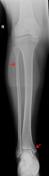
Image 4 CT (3D-VR-bone window) ( update )
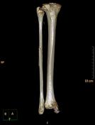
Image 5 X-ray (Frontal) ( update )
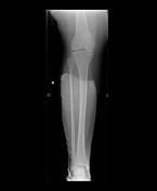
Image 6 X-ray (Frontal) ( update )

Image 7 X-ray (Frontal) ( update )
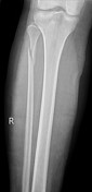
Image 8 X-ray (Lateral) ( update )


 Unable to process the form. Check for errors and try again.