Spondylolysis
Updates to Article Attributes
Spondylolysis is a defect in the pars interarticularis of the neural arch, the portion of the neural arch that connects the superior and inferior articular facets. It is commonly known as pars interarticularis defect or more simply as pars defect.
Epidemiology
Spondylolysis is present in ~5% of the population 2 and higher in the adolescent athletic population. It is more common in men than in women 1.
Associations
~65% of patients with spondylolysis will progress to spondylolisthesis 2, which is seen radiographically in ~25% 4; in most patients this occurs before the age of 16
idiopathic scoliosis: ~5% 8
Clinical presentation
Spondylolysis is commonly asymptomatic. Symptomatic patients often have pain with extension and/or rotation of the lumbar spine. Approximately 25% of individuals with spondylolysis have symptoms at some time.
It is a common cause of low back pain in adolescents and may be the cause of low back pain in ~50% of adolescent athletes 7.
Pathology
Spondylolysis may be developmental or acquired. Developmental defects may develop in patients <10 years 7. Acquired defects have two main mechanisms:
repeated microtrauma, resulting in a stress injury and eventual fracture of the pars interarticularis; a dysplastic pars is usually present
traumatic pars defects result from high-energy trauma where there is hyperextension of the lumbar spine and are rare in a congenitally normal vertebra
Location
~90% of cases of spondylolysis occur at the L5 level and ~10% occur at the L4 level 1,2
unilateral or bilateral
Radiographic features
Imaging features depend on the age of the lesion. CT is considered the gold standard although MRI should be used as the first-line imaging modality in adolescents 7.
Plain radiograph
limited sensitivity compared to SPECT and CT 4
Scotty dog sign: on oblique radiographs, a break in the pars interarticularis can have the appearance of a collar around the dog's neck
CT/MRI
wide-canal sign may be present on sagittal images when there is spondylolisthesis 3
deviation of the spinous process
sclerosis of the contralateral pedicle
Hollenberg classification
This is a classification system mostly based on MRI features but correlates well with SPECT-CT 6,7:
grade 0: normal pars interarticularis; MRI: no signal abnormality, pars interarticularis intact
grade I: stress reaction; MRI: marrow oedema; intact cortical margins
grade II: incomplete stress fracture; MRI: marrow oedema; incomplete cortical fracture or fissure
grade III: acute complete stress fracture; MRI: marrow oedema; complete cortical fracture extending through pars interarticularis
grade IV: chronic stress fracture; MRI: no marrow oedema; fractures completely extending through pars interarticularis
Treatment and prognosis
Surgery is only considered in rare circumstances as most cases respond to conservative management 2. Incomplete fractures demonstrate good healing rates with conservative management 7.
Differential diagnosis
-<p><strong>Spondylolysis</strong> is a defect in the <a href="/articles/pars-interarticularis">pars interarticularis</a> of the neural arch, the portion of the neural arch that connects the superior and inferior articular facets. It is commonly known as<strong> pars interarticularis defect</strong> or more simply as <strong>pars defect</strong>. </p><h4>Epidemiology</h4><p>Spondylolysis is present in ~5% of the population <sup>2</sup> and higher in the adolescent athletic population. It is more common in men than in women<sup> 1</sup>.</p><h5>Associations</h5><ul>-<li><p>~65% of patients with spondylolysis will progress to <a href="/articles/spondylolisthesis-1">spondylolisthesis</a> <sup>2</sup>, which is seen radiographically in ~25% <sup>4</sup>; in most patients this occurs before the age of 16</p></li>-<li><p><a href="/articles/spina-bifida-occulta">spina bifida occulta</a></p></li>-<li><p>idiopathic <a href="/articles/scoliosis">scoliosis</a>: ~5% <sup>8</sup></p></li>-</ul><h4>Clinical presentation</h4><p>Spondylolysis is commonly asymptomatic. Symptomatic patients often have pain with extension and/or rotation of the lumbar spine. Approximately 25% of individuals with spondylolysis have symptoms at some time.</p><p>It is a common cause of <a href="/articles/low-back-pain">low back pain</a> in adolescents and may be the cause of low back pain in ~50% of adolescent athletes <sup>7</sup>.</p><h4>Pathology</h4><p>Spondylolysis may be developmental or acquired. Developmental defects may develop in patients <10 years <sup>7</sup>. Acquired defects have two main mechanisms:</p><ul>-<li><p>repeated microtrauma, resulting in a <a href="/articles/stress-fracture-2">stress injury</a> and eventual fracture of the pars interarticularis; a dysplastic pars is usually present</p></li>-<li><p>traumatic pars defects result from high-energy trauma where there is hyperextension of the lumbar spine and are rare in a congenitally normal vertebra</p></li>-</ul><h5>Location</h5><ul>-<li><p>~90% of cases of spondylolysis occur at the L5 level and ~10% occur at the L4 level <sup>1,2</sup></p></li>-<li><p>unilateral or bilateral</p></li>-</ul><h4>Radiographic features</h4><p>Imaging features depend on the age of the lesion. CT is considered the gold standard although MRI should be used as the first-line imaging modality in adolescents <sup>7</sup>.</p><h5>Plain radiograph</h5><ul>-<li><p>limited sensitivity compared to SPECT and CT <sup>4</sup></p></li>-<li><p><a href="/articles/scottie-dog-sign-spine">Scotty dog sign</a>: on oblique radiographs, a break in the pars interarticularis can have the appearance of a collar around the dog's neck</p></li>-<li><p><a href="/articles/inverted-napoleon-hat-sign">inverted Napoleon hat sign</a></p></li>-</ul><h5>CT/MRI</h5><ul>-<li><p><a href="/articles/wide-canal-sign">wide-canal sign</a> may be present on sagittal images when there is spondylolisthesis <sup>3</sup></p></li>-<li><p>deviation of the spinous process</p></li>-<li><p>sclerosis of the contralateral pedicle</p></li>-</ul><h5>Hollenberg classification</h5><p>This is a classification system mostly based on MRI features but correlates well with SPECT-CT <sup>6,7</sup>:</p><ul>-<li><p><strong>grade 0:</strong> normal pars interarticularis; MRI: no signal abnormality, pars interarticularis intact</p></li>-<li><p><strong>grade I:</strong> stress reaction; MRI: marrow oedema; intact cortical margins</p></li>-<li><p><strong>grade II:</strong> incomplete stress fracture; MRI: marrow oedema; incomplete cortical fracture or fissure</p></li>-<li><p><strong>grade III: </strong>acute complete stress fracture; MRI: marrow oedema; complete cortical fracture extending through pars interarticularis</p></li>-<li><p><strong>grade IV: </strong>chronic stress fracture; MRI: no marrow oedema; fractures completely extending through pars interarticularis</p></li>-</ul><h4>Treatment and prognosis</h4><p>Surgery is only considered in rare circumstances as most cases respond to conservative management <sup>2</sup>. Incomplete fractures demonstrate good healing rates with conservative management <sup>7</sup>.</p><h4>Differential diagnosis</h4><ul>-<li><p><a href="/articles/osteoid-osteoma">osteoid osteoma</a></p></li>-<li><p><a href="/articles/osteomyelitis">osteomyelitis</a></p></li>-<li><p><a href="/articles/retroisthmic-cleft" title="Retroisthmic cleft">retroisthmic cleft</a></p></li>- +<p><strong>Spondylolysis</strong> is a defect in the <a href="/articles/pars-interarticularis">pars interarticularis</a> of the neural arch, the portion of the neural arch that connects the superior and inferior articular facets. It is commonly known as<strong> pars interarticularis defect</strong> or more simply as <strong>pars defect</strong>. </p><h4>Epidemiology</h4><p>Spondylolysis is present in ~5% of the population <sup>2</sup> and higher in the adolescent athletic population. It is more common in men than in women<sup> 1</sup>.</p><h5>Associations</h5><ul>
- +<li><p>~65% of patients with spondylolysis will progress to <a href="/articles/spondylolisthesis-1">spondylolisthesis</a> <sup>2</sup>, which is seen radiographically in ~25% <sup>4</sup>; in most patients this occurs before the age of 16</p></li>
- +<li><p><a href="/articles/spina-bifida-occulta">spina bifida occulta</a></p></li>
- +<li><p>idiopathic <a href="/articles/scoliosis">scoliosis</a>: ~5% <sup>8</sup></p></li>
- +</ul><h4>Clinical presentation</h4><p>Spondylolysis is commonly asymptomatic. Symptomatic patients often have pain with extension and/or rotation of the lumbar spine. Approximately 25% of individuals with spondylolysis have symptoms at some time.</p><p>It is a common cause of <a href="/articles/low-back-pain">low back pain</a> in adolescents and may be the cause of low back pain in ~50% of adolescent athletes <sup>7</sup>.</p><h4>Pathology</h4><p>Spondylolysis may be developmental or acquired. Developmental defects may develop in patients <10 years <sup>7</sup>. Acquired defects have two main mechanisms:</p><ul>
- +<li><p>repeated microtrauma, resulting in a <a href="/articles/stress-fracture-2">stress injury</a> and eventual fracture of the pars interarticularis; a dysplastic pars is usually present</p></li>
- +<li><p>traumatic pars defects result from high-energy trauma where there is hyperextension of the lumbar spine and are rare in a congenitally normal vertebra</p></li>
- +</ul><h5>Location</h5><ul>
- +<li><p>~90% of cases of spondylolysis occur at the L5 level and ~10% occur at the L4 level <sup>1,2</sup></p></li>
- +<li><p>unilateral or bilateral</p></li>
- +</ul><h4>Radiographic features</h4><p>Imaging features depend on the age of the lesion. CT is considered the gold standard although MRI should be used as the first-line imaging modality in adolescents <sup>7</sup>.</p><h5>Plain radiograph</h5><ul>
- +<li><p>limited sensitivity compared to SPECT and CT <sup>4</sup></p></li>
- +<li><p><a href="/articles/scottie-dog-sign-spine">Scotty dog sign</a>: on oblique radiographs, a break in the pars interarticularis can have the appearance of a collar around the dog's neck</p></li>
- +<li><p><a href="/articles/inverted-napoleon-hat-sign">inverted Napoleon hat sign</a></p></li>
- +</ul><h5>CT/MRI</h5><ul>
- +<li><p><a href="/articles/wide-canal-sign">wide-canal sign</a> may be present on sagittal images when there is spondylolisthesis <sup>3</sup></p></li>
- +<li><p>deviation of the spinous process</p></li>
- +<li><p>sclerosis of the contralateral pedicle</p></li>
- +</ul><h5>Hollenberg classification</h5><p>This is a classification system mostly based on MRI features but correlates well with SPECT-CT <sup>6,7</sup>:</p><ul>
- +<li><p><strong>grade 0:</strong> normal pars interarticularis; MRI: no signal abnormality, pars interarticularis intact</p></li>
- +<li><p><strong>grade I:</strong> stress reaction; MRI: marrow oedema; intact cortical margins</p></li>
- +<li><p><strong>grade II:</strong> incomplete stress fracture; MRI: marrow oedema; incomplete cortical fracture or fissure</p></li>
- +<li><p><strong>grade III: </strong>acute complete stress fracture; MRI: marrow oedema; complete cortical fracture extending through pars interarticularis</p></li>
- +<li><p><strong>grade IV: </strong>chronic stress fracture; MRI: no marrow oedema; fractures completely extending through pars interarticularis</p></li>
- +</ul><h4>Treatment and prognosis</h4><p>Surgery is only considered in rare circumstances as most cases respond to conservative management <sup>2</sup>. Incomplete fractures demonstrate good healing rates with conservative management <sup>7</sup>.</p><h4>Differential diagnosis</h4><ul>
- +<li><p><a href="/articles/osteoid-osteoma">osteoid osteoma</a></p></li>
- +<li><p><a href="/articles/osteomyelitis">osteomyelitis</a></p></li>
- +<li><p><a href="/articles/retroisthmic-cleft" title="Retroisthmic cleft">retroisthmic cleft</a></p></li>
Image ( destroy )
Image 1 X-ray (Lateral) ( update )

Image 2 Annotated image (sagittal bone window) ( update )

Image 3 CT (MIP) ( update )
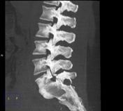
Image 4 CT (bone window) ( update )
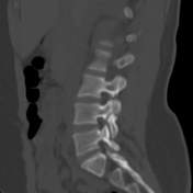
Image 5 X-ray (Lateral) ( update )
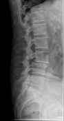
Image 6 Annotated image (Sagittal T2) ( update )

Image 7 CT (bone window) ( update )
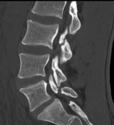
Image 8 MRI (T1 VIBE) ( update )

Image 9 X-ray (Lateral) ( update )

Image 10 X-ray (Oblique- zoomed) ( create )


 Unable to process the form. Check for errors and try again.