
Xanthogranulomatous pyelonephritis (XGP) is a rare form of chronic pyelonephritis and represents a chronic granulomatous disease resulting in a non-functioning kidney. Radiographic features are usually specific.
On this page:
Epidemiology
Xanthogranulomatous pyelonephritis is seen essentially in all age groups, but most frequently presents in middle-aged to elderly patients 1,5. There is a 2:1 female predilection, presumably relating to an increased incidence of urinary tract infections and thus struvite (staghorn) calculi. There is also an increased incidence in patients with diabetes mellitus.
Clinical presentation
The clinical presentation is typically vague, consisting of constitutional symptoms such as malaise, weight loss and low-grade fever. Hematuria and flank pain are sometimes encountered 4.
Despite often absent urinary tract symptoms, pyuria and positive urinary cultures are present in the majority of cases (95% and 60% respectively) 2.
Pathology
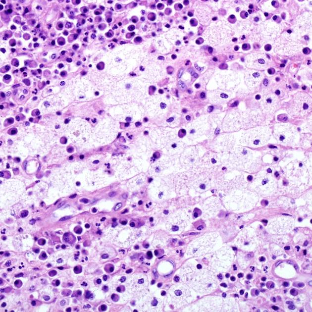
Xanthogranulomatous pyelonephritis is, as the name suggests, a chronic granulomatous process believed to be the result of subacute/chronic infection inciting a chronic but incomplete immune reaction 1,4. Various bacteria are isolated, however, the most commonly isolated species are Escherichia coli and Proteus mirabilis 1,4.
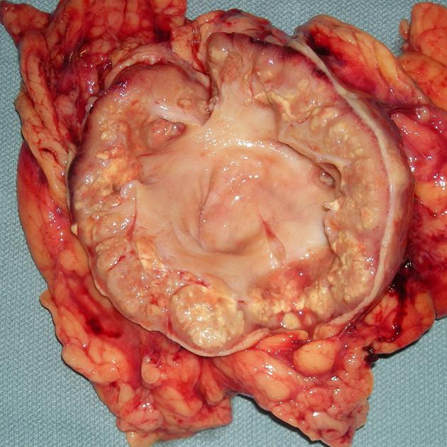
The kidney is eventually replaced by a mass of reactive tissue, surrounding the usually present (90%) inciting staghorn calculus with associated hydronephrosis of a greater or lesser degree. Foamy (lipid-laden) macrophages predominate 1,4.
The inflammatory process eventually extends into the perinephric tissues and even adjacent organs 5.
Staging
One method of staging (originally proposed by Malek et al.) is based on the degree of involvement of the adjacent tissues 6:
stage I: the disease is confined to the renal parenchyma only
stage II: involves renal parenchyma as well as an extension to perirenal/perinephric fat
stage III: the disease extends into the perirenal and pararenal spaces or diffuse retroperitoneum
This staging was originally described in a pediatric population but can be applied to adults.
Radiographic features
Two forms of the disease are recognized both macroscopically and on imaging 1,5:
diffuse (90%)
-
focal/tumefactive (10%)
sometimes a truly focal process in a normal kidney
in other instances, this represents diffuse xanthogranulomatous pyelonephritis of one moiety of a duplex system
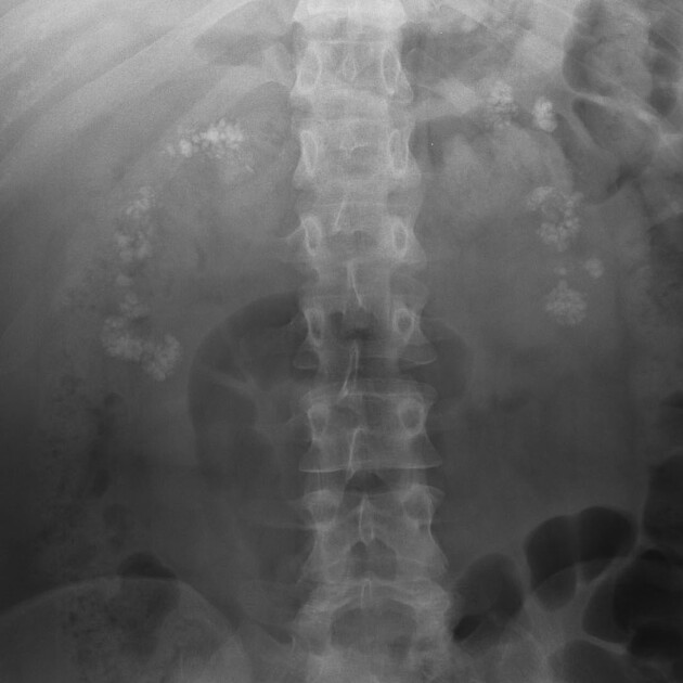
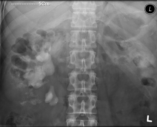
Plain radiograph
Plain radiograph findings are difficult to distinguish from a routine staghorn calculus, although fragmentation and enlargement of the renal outline may be seen. A calculus is not always present; in such cases, it is not possible to make a plain film diagnosis.
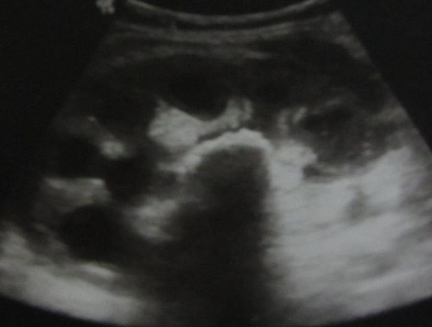
Ultrasound
Ultrasound examination demonstrates an enlarged and distorted renal outline, with loss of the normal renal architecture and (usually) a centrally-located shadowing calculus.
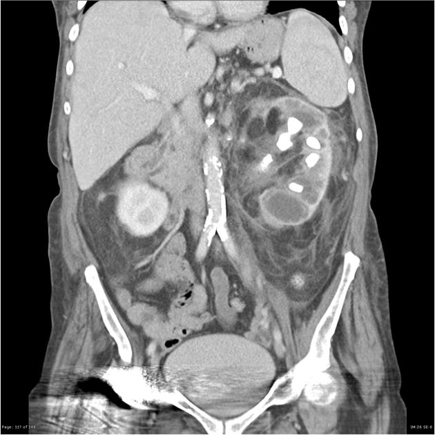
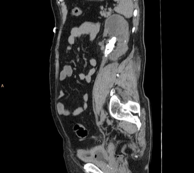
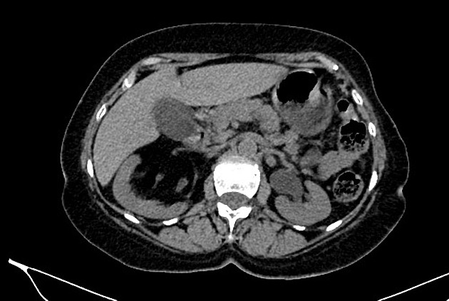
CT
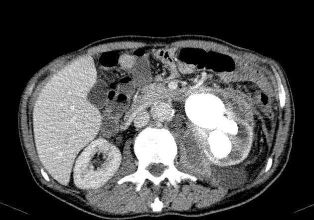
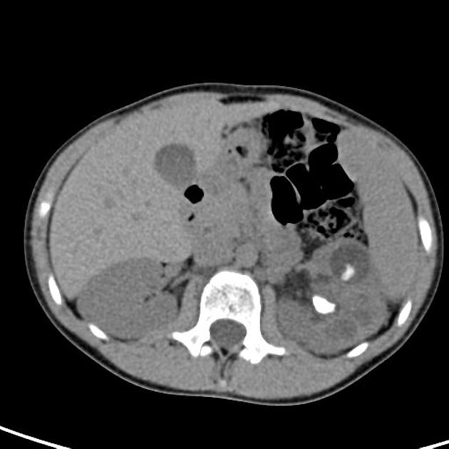
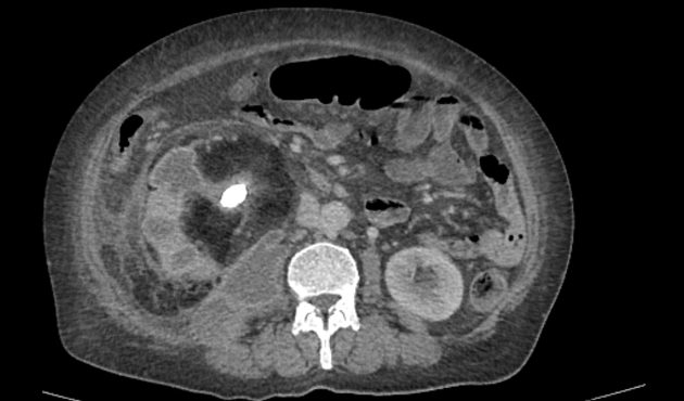
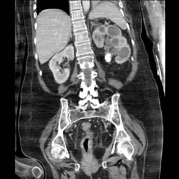
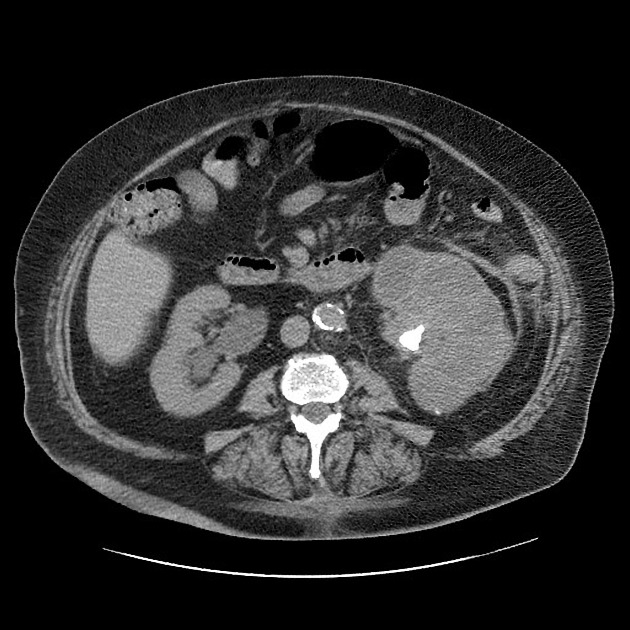
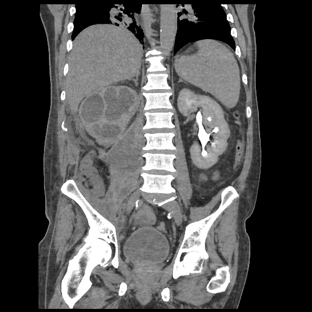
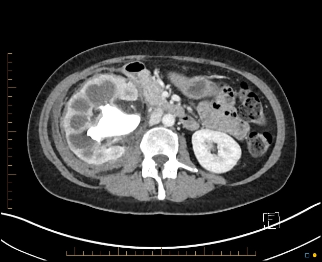
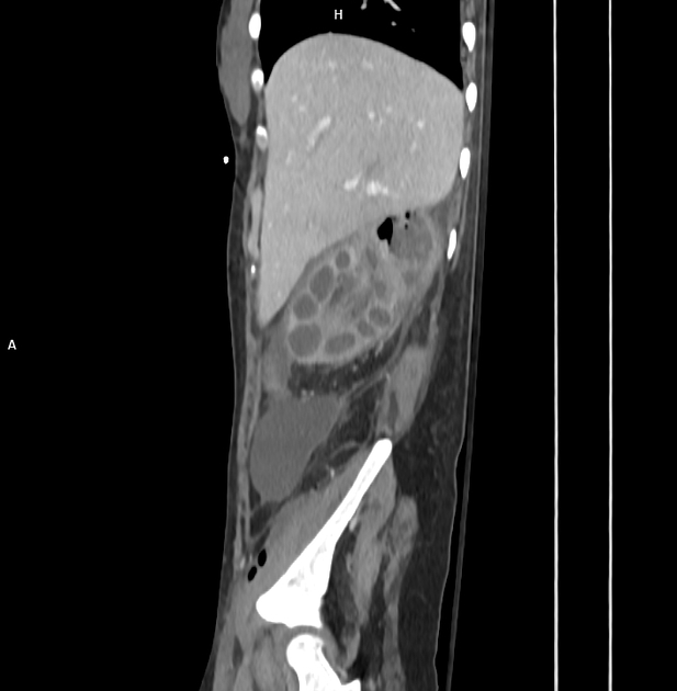
CT findings are most helpful in reaching the correct diagnosis. The normal renal outline is lost and enlarged with a paradoxical contracted renal pelvis. The calyces, in contrast, are dilated giving a multiloculated appearance that has been likened to the paw print of a bear (bear's paw sign) 3. Sometimes there is a perinephric extension with thickening of Gerota's fascia. Calcification and a staghorn calculus can be better delineated on CT scan.
There is also a more focal form of xanthogranulomatous pyelonephritis, where a small low attenuation mass with an associated calculus is seen adjacent to a calyx or in one pole of a kidney.
Urography (CT/conventional)
In most cases, there is little, if any, renal function in the affected kidney 1.
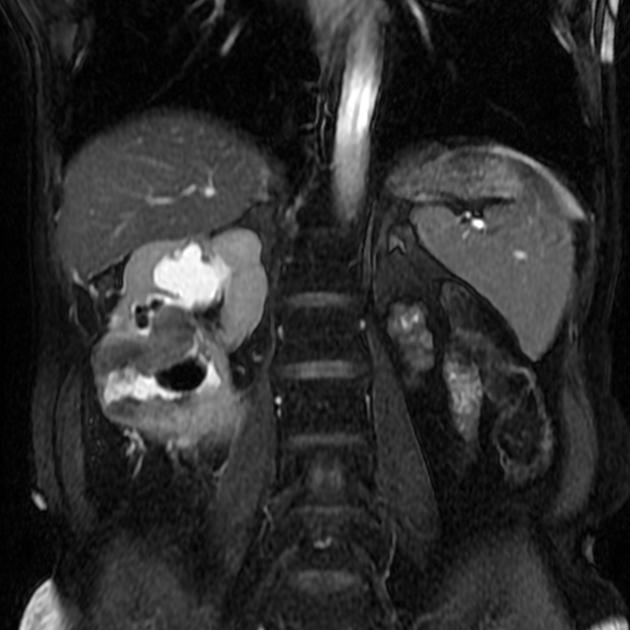
MRI
MRI appearances mirror the heterogeneous nature of the mass with solid and cystic components surrounding a central staghorn calculus. As such, the signal is heterogeneous on all sequences.
Treatment and prognosis
If xanthogranulomatous pyelonephritis becomes established, no conservative or medical therapies exist. Surgical nephrectomy is usually curative 4,5. The presence of an inflammatory reaction in adjacent tissues often requires a large operative field and an anterolateral transperitoneal approach 5.
Differential diagnosis
The differential is narrow when the entire kidney is affected and cross-sectional imaging has been obtained and is largely limited to renal tuberculosis, however, this usually results in a shrunken calcified putty kidney.
In cases where typical features are not present (e.g. no staghorn calculus, focal disease only) then other entities to be considered include:


 Unable to process the form. Check for errors and try again.