
Presentation
Acute abdominal pain and vomiting.
Patient Data
Age: 50 years
Gender: Male
From the case:
Acute appendicitis


Download
Info

Delayed phase MDCT reveals an enlarged appendix, measuring about 15 mm in diameter, with enhancing, thickened wall and surrounding stranding. A high density calculus (appendicolith) is noted within.
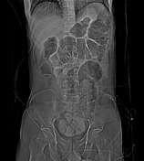
Three calcified fecaliths are visible even in topogram.


 Unable to process the form. Check for errors and try again.