
Presentation
Non-descriptive visceral pain in the periumbilical region, followed by nausea. Previous radiography study was unremarkable. Previous US study showed signs of inflamed mesenteric fat in the right lower quadrant.
Patient Data
Note: This case has been tagged as "legacy" as it no longer meets image preparation and/or other case publication guidelines.


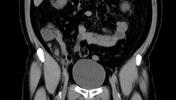
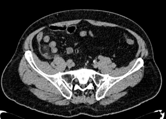
Right lower quadrant: dilated appendix with a small appendicolith and surrounding inflammatory stranding.
Case Discussion
The prevalence of appendicitis in patients who present with abdominal pain to the emergency department is approximately 14%.
Traditionally, acute appendicitis has been diagnosed by clinical findings.
CT is highly sensitive (94-98%) and specific (up to 97%) for the diagnosis of acute appendicitis, and allows for alternative causes of abdominal pain also to be diagnosed. In this case, a non-contrast study, as institution protocol, was enough to determine the diagnosis.


 Unable to process the form. Check for errors and try again.