
Presentation
Abdominal pain
Patient Data
Age: 40 years
Gender: Female


Download
Info
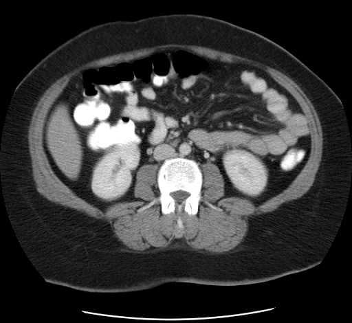
Dilated appendix with significant surrounding stranding. However, no drainable collection or extraluminal gas can be seen.
The right ovarian vein is distended and has a thick enhancing wall, along with hypodensity in the lumen extending to the middle portion of the vein (the lumen is not enhancing vs the contralateral left ovarian vein). The changes of the right ovarian vein are better appreciated on the coronal images.
No abdominal free fluid.
No retroperitoneal or pelvic enlarged lymph nodes.
Interpretation: Features of acute appendicitis complicated by ovarian vein thrombosis.


 Unable to process the form. Check for errors and try again.