
Presentation
Right flank pain. Renal colic?
Patient Data
Age: 30 years
Gender: Male


Download
Info
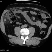
Non contrast CT KUB obtained to evaluate for renal stones demonstrates the appendix, located behind and lateral to the cecum (paracecal), with evidence of acute inflammation and a luminal fecolith at its base. No renal or ureteric calculi. No free gas.

Download
Info
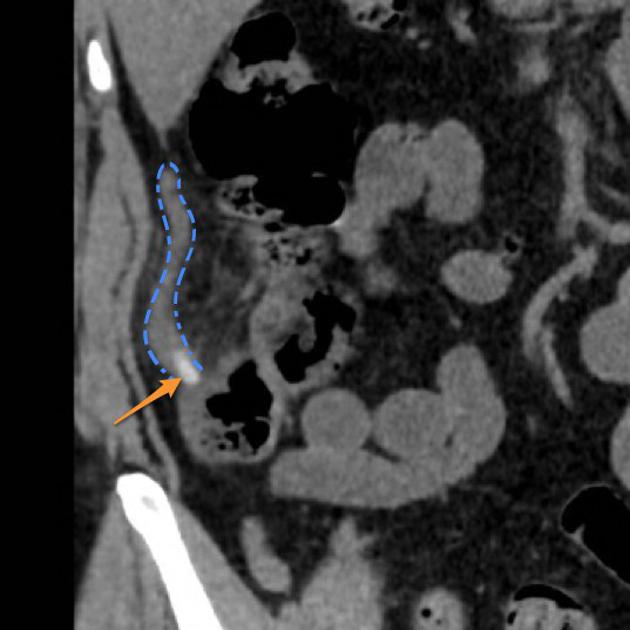
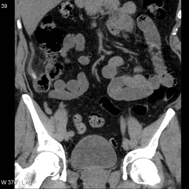
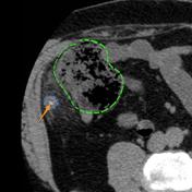
Coronal and axial images demonstrate the appendix (blue dotted line) to be located to lateral to the cecum (green dotted line) with extensive surrounding fat stranding. At the neck of the appendix is a calcific density consistent with an appendicolith (orange arrow).
Case Discussion
This case illustrates how a less common location of the appendix (in this case paracecal) can result in atypical presentation which can mimic other pathologies.


 Unable to process the form. Check for errors and try again.