
Presentation
Right sided weakness.
Patient Data
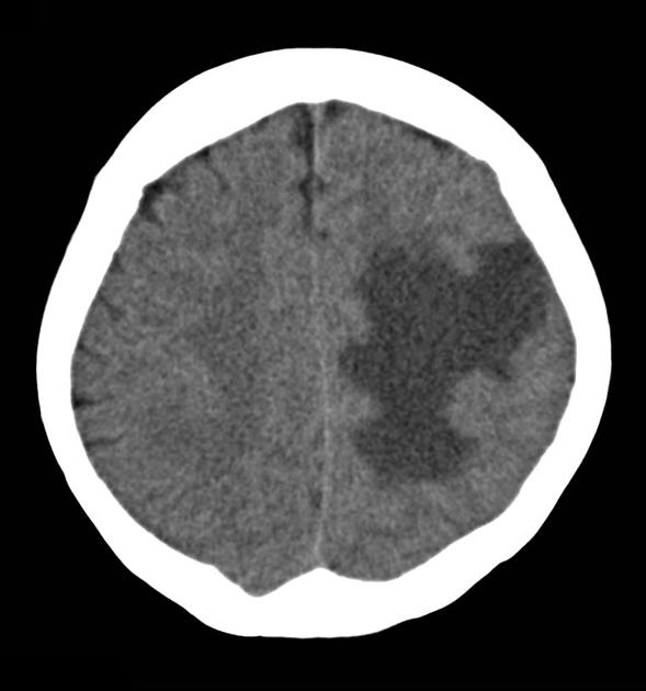
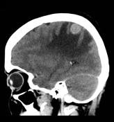
A 2cm rounded mass is present in the post-central gyrus, which is iso-dense to cortex pre-contrast and demonstrates homogeneous contrast enhancement. It is located at the grey-white matter interface and is surrounded by extensive vasogenic edema, which exerts significant mass effect.


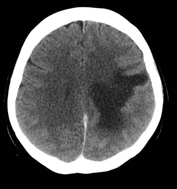
Following the administration of contrast the mass demonstrates relatively vivid enhancement. It remains an isolated abnormality.
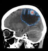
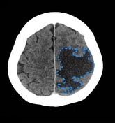

The enhancing mass ( * ) is surrounded by vasogenic edema (blue dotted line) which is confined to the white matter.
Case Discussion
Although typically we think of multiplicity as being the hallmark of cerebral metastases, a large number are seemingly solitary at the time of diagnosis. Thus metastatic disease needs to be entertained in the differential of a solitary cerebral mass.
In this case the patient had a known history of metastatic colorectal carcinoma.


 Unable to process the form. Check for errors and try again.