
Charcot joint - spine
Diagnosis almost certain
Updates to Study Attributes
Findings
was changed:
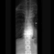
A focal thoracic kyphosis is present due to severe destructure change involving adjacent vertebra with anterolisthesis.
Images Changes:
Image X-ray (Lateral) ( update )

Description
was removed:
Image X-ray (Frontal) ( update )
Description
was removed:
Updates to Study Attributes
Findings
was changed:
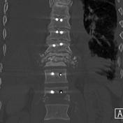
The patient went on to have surgical fusion and biopsy.
Images Changes:
Image CT (bone window) ( update )

Description
was removed:
Image CT (bone window) ( update )
Description
was removed:
Updates to Case Attributes
Body
was changed:
PathThis is a case of histologically proven Charcot joint in a diabetic patient. Note the distinctive difference in bone density above and below the Charcot joint (sclerotic above and lucent below) joint on plain radiographs.
-<p>Path proven <a href="/articles/charcot-joint">Charcot joint</a> in a diabetic patient. Note the distinctive difference in bone density above and below the Charcot joint (sclerotic above and lucent below). </p>- +<p>This is a case of histologically proven <a href="/articles/charcot-joint">Charcot joint</a> in a diabetic patient. Note the distinctive difference in bone density above and below the Charcot joint on plain radiographs. </p>
Age
was set to
Elderly. .
Gender
was set to
Female.


 Unable to process the form. Check for errors and try again.