
Patient Data
Age: 6 years
Gender: Male
Note: This case has been tagged as "legacy" as it no longer meets image preparation and/or other case publication guidelines.
From the case:
Extradural hematoma


Download
Info
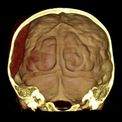
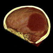
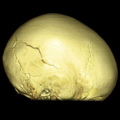
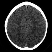
A right-sided parietal fracture with underlying bi-convex (lentiform) extradural hematoma. Note how the posterior edge of the collection is bounded by the parieto-occipital suture.

Download
Info

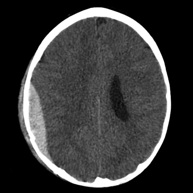
The patient was treated conservatively. At 3 month follow-up, the extradural hematoma had essentially resolved with minor dural calcification remaining.
Case Discussion
Not all extradural hematomas need surgical management. This is particularly true of smaller venous hematomas (e.g. those located in the anterior middle cranial fossa).
Naturally very close clinical and imaging follow-up is required particularly in the first few days.


 Unable to process the form. Check for errors and try again.