
Presentation
Chronic left knee pain lasting for almost a year without previous trauma or signs of inflammation. She was referred for a routine MRI of the left knee to rule out internal derangement.
Patient Data

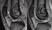
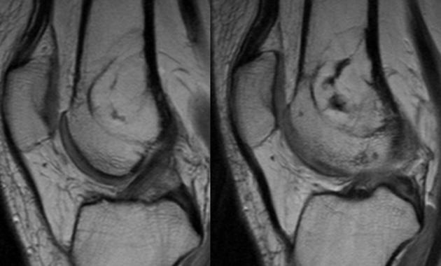
Sagittal T1-weighted images show a lesion in the distal femur, that is isointense to fat marrow and contains bony inclusions. It has a sharp, signal-free border to the normal bone.
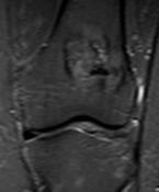
T2*-weighting shows bone susceptibility effects within and around the lesion. In Coronal STIR, most of the lesion's signal is suppressed, except for some hyperintense patches.
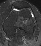
Transverse PD-FSE fat sat, the predominantly fatty components of the lesion appear dark. There is, however, some focal bright signal.
Case Discussion
Plain films had not been obtained prior to the MRI examination. T1-weighted MRI and T2* GRE showed a bright lesion with sclerotic wall thickening and bony septa. On STIR and fat-suppressed, PD-weighted images, most of the interior signal of the lesion was suppressed, except for some irregular hyperintense inclusions. The lesion showed no signs of aggressive behavior. Intravenous contrast medium was not administered.


 Unable to process the form. Check for errors and try again.