
Presentation
Short of breath
Patient Data
Note: This case has been tagged as "legacy" as it no longer meets image preparation and/or other case publication guidelines.

Chest x-ray (slightly rotated to the right) demonstrates a large opacity obliterating the left hilum. It is associated with some volume loss in the left hemithorax and what appears to be a pleural effusion.
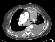

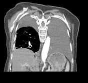
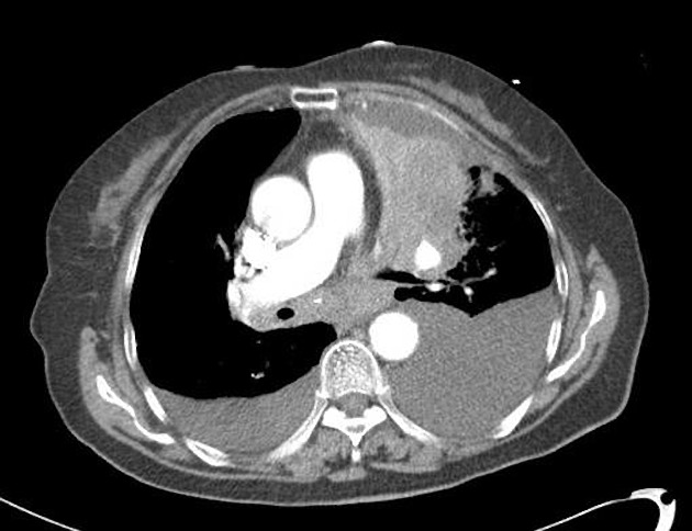
Selected images from a CT of the chest and upper abdomen confirm a large mass occupying the majority of the left upper lobe, with distal collapse. A large left and small right pleural effusion are also present with resulting dependent atelectasis of parts of the lower lobe.
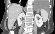
Mediastinal nodal enlargement is present. The adrenal glands do not appear clearly abnormal on the images provided.
Case Discussion
A left upper lobe lung primary carcinoma (squamous cell carcinoma) was confirmed histologically.


 Unable to process the form. Check for errors and try again.