
Presentation
History of treated pulmonary tuberculosis, chronic smoking.
Patient Data

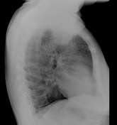
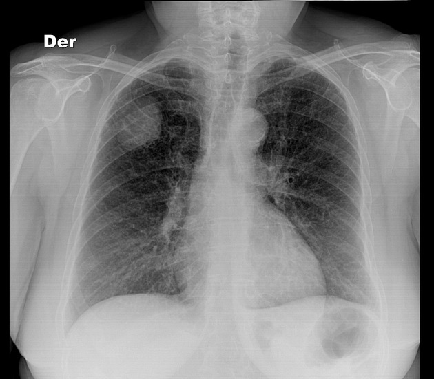
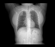
There are diffuse reticular pattern. In the periphery of the right upper lobe, there is a 3cm nodular opacity with quite well defined contours. Prominence of the aortic knob, shows calcified atheromas. No signs of pleural effusion or pneumothorax. Dorsal spondylosis.
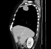

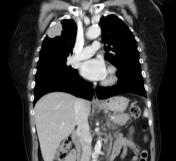
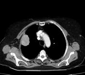
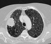

CT confirms a subpleural ovoid mass, measuring 4.6 x 3.6 cm, that enhances with contrast medium but no signs of local infiltration. A second smaller nodule is seen in the inferior lingula segment.
Background of emphysema and reticular changes.
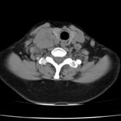
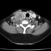
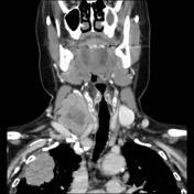
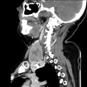
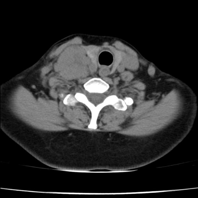
Large nodal mass with central necrosis, which compresses the ipsilateral internal jugular vein without occluding it. A solitary hypodense rounded nodule is seen in the right thyroid lobe.
Case Discussion
This case is a good example of a lung tumor with neck metastasis.
Histological confirmation for management and treatment planning is essential. This is an excellent example of where a less invasive and lower risk tissue diagnosis can be ascertained by a ultrasound guided neck node biopsy, rather than a CT guided lung biopsy.


 Unable to process the form. Check for errors and try again.