
Presentation
No history available.
Patient Data
Age: Young adult
Gender: Female
Download
Info
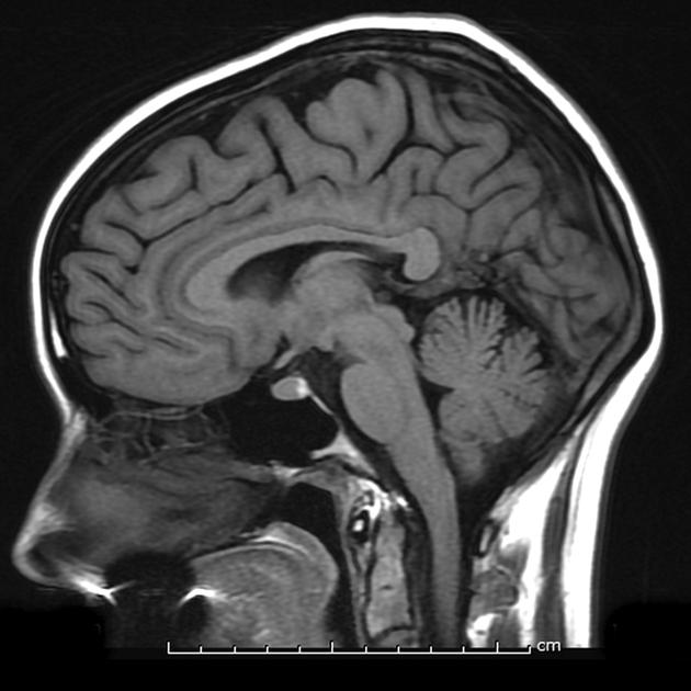
Single midline sagittal T1 weighted image demonstrates normal anatomy, including the posterior pituitary bright spot.
Case Discussion
Normal midline brain MRI, with a normal posterior pituitary bright spot.
For similar examples, please refer to examples of normal brain imaging.


 Unable to process the form. Check for errors and try again.