
Presentation
A known case of peripheral primitive neuroectodermal tumor referred to our hospital for total hip arthroplasty
Patient Data
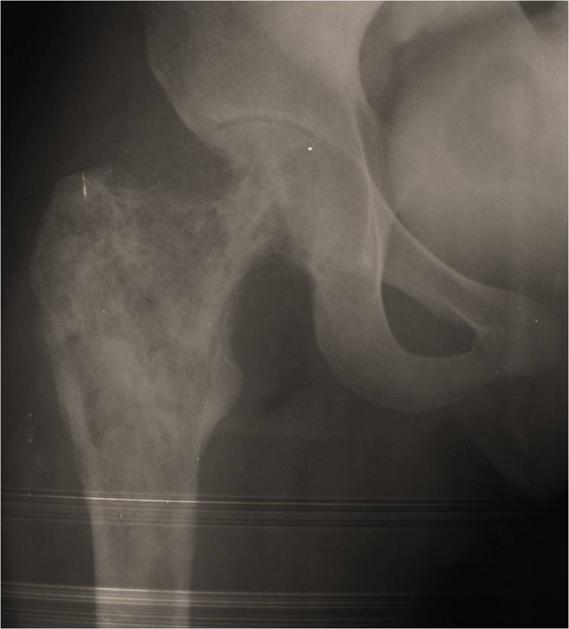
An ill-defined lytic lesion with a moth-eaten appearance is seen in the metaphysis, neck and head of the right femur.
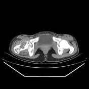

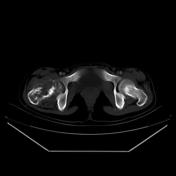

Patchy destructive bony changes associated with soft tissue components demonstrated within the femoral head and neck as well as amorphous periosteal reaction. There is also evidence of bone marrow involvement. The tumor has grown along a needle biopsy track in the subcutaneous tissue
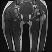
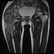
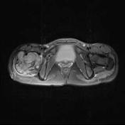
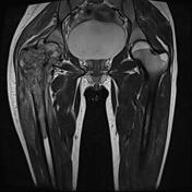
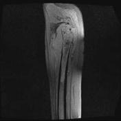
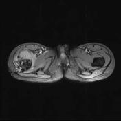

MRI shows bone marrow involvement and soft tissue component of the tumor which surround the femur. The soft tissue mass signal is similar to that of muscle on T1WI and high on T2WI.
Following the administration of gadolinium, the mass shows intense enhancement.
Case Discussion
Peripheral primitive neuroectodermal tumor (pPNET) of the proximal right femur in a 20 year old female.


 Unable to process the form. Check for errors and try again.