
Portal vein anatomy
Diagnosis not applicable
Updates to Study Attributes
Modality
changed from CT to Annotated image.
Images Changes:
Image 1 Annotated image (Coronal) ( update )
Position
was set to
.
Image 2 Annotated image (Coronal) ( update )

Position
was set to
.
Image 3 Annotated image (Coronal) ( update )

Position
was set to
.
Image 4 Annotated image (Axial) ( update )
Position
was set to
.
Image 5 Annotated image (Axial) ( update )

Position
was set to
.
Updates to Case Attributes
Body
was changed:
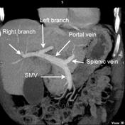
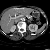
The portal vein is formed by the union of the SMV (superior mesenteric vein) with the splenic vein.
The final image is a wedge portal venogram obtained during a TIPS (transjugular intrahepatic portosytemic shunt) procedure, using using carbon dioxide as a contrast agent. The venogram demonstrates a rather prominent left gastric vein (aka coronary vein (a.k.a. coronary vein) draining into the SMV-Splenic-splenic venous confluence. This patient was known to known to have portal hypertension which probably accounts for the size of this vein.
-<p>The <a href="/articles/portal-vein" title="Portal vein">portal vein</a> is formed by the union of the SMV (superior mesenteric vein) with the splenic vein. </p><p>The final image is a wedge portal venogram obtained during a TIPS (transjugular intrahepatic portosytemic shunt) procedure, using carbon dioxide as a contrast agent</span>. The venogram demonstrates a rather prominent left gastric vein (aka coronary vein) draining into the SMV-Splenic venous confluence. This patient was known to have portal hypertension which probably accounts for the size of this vein.</p>- +<p>The <a href="/articles/portal-vein">portal vein</a> is formed by the union of the <a title="Superior mesenteric vein (SMV)" href="/articles/superior-mesenteric-vein">SMV (superior mesenteric vein)</a> with the <a title="Splenic vein" href="/articles/splenic-vein">splenic vein</a>. </p><p>The final image is a wedge portal venogram obtained during a <a title="TIPS" href="/articles/transjugular-intrahepatic-portosystemic-shunt">TIPS</a> (transjugular intrahepatic portosytemic shunt) procedure, using carbon dioxide as a contrast agent. The venogram demonstrates a rather prominent <a title="Left gastric vein" href="/articles/left-gastric-vein">left gastric vein</a> (a.k.a. coronary vein) draining into the SMV-splenic venous confluence. This patient was known to have portal hypertension which probably accounts for the size of this vein.</p>
Systems changed:
- Hepatobiliary


 Unable to process the form. Check for errors and try again.