Presentation
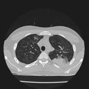
Investigation of left upper lobe consolidation
Patient Data
Age: 18
Gender: Male
![](https://prod-images-static.radiopaedia.org/images/4984024/a38100df04e96e30a1206fb1dcc448_thumb.jpg)
![This study is a stack](/packs/stack-YQKLCKBI.gif)
![](https://prod-images-static.radiopaedia.org/images/4982685/0b6d4544a41b2e5e2039fe9de71a9a_thumb.jpg)
![This study is a stack](/packs/stack-YQKLCKBI.gif)
![](https://prod-images-static.radiopaedia.org/images/4983350/651507e39dfccbee5eb59540e9d7a9_thumb.jpg)
![This study is a stack](/packs/stack-YQKLCKBI.gif)
![](https://prod-images-static.radiopaedia.org/images/4983024/3ec621ddfde10e0eeaaf4449fd2627_thumb.jpg)
![This study is a stack](/packs/stack-YQKLCKBI.gif)
![](https://prod-images-static.radiopaedia.org/images/4983789/21d86069031b43a7f0e3c1d93b7369_thumb.jpg)
![This study is a stack](/packs/stack-YQKLCKBI.gif)
Download
Info
![](https://prod-images-static.radiopaedia.org/images/4984024/a38100df04e96e30a1206fb1dcc448_big_gallery.jpg)
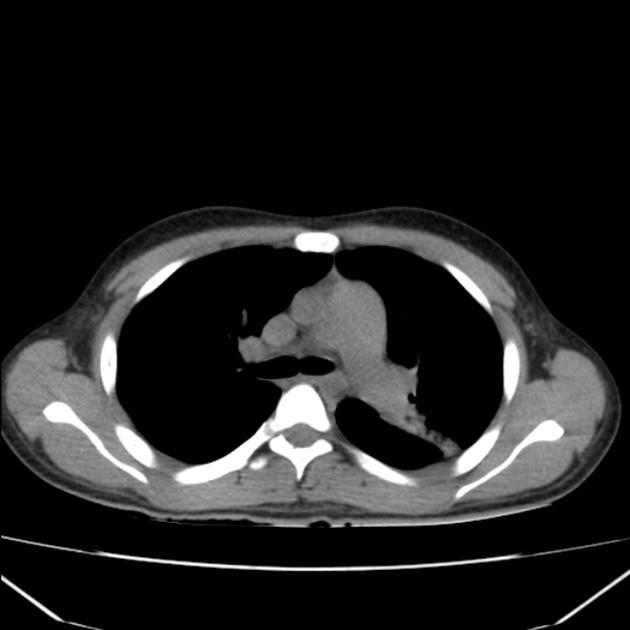
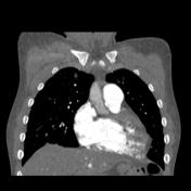
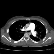
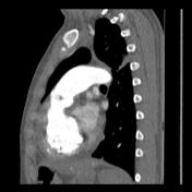
There is marked enlargement of the main pulmonary artery and is wider than the aorta on the CT. Signs of right heart ventricle overload also shown; There is also a small concurrent left upper lobe pneumonia
Case Discussion
Features of pulmonary hypertension