Patient Data

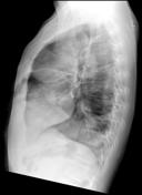
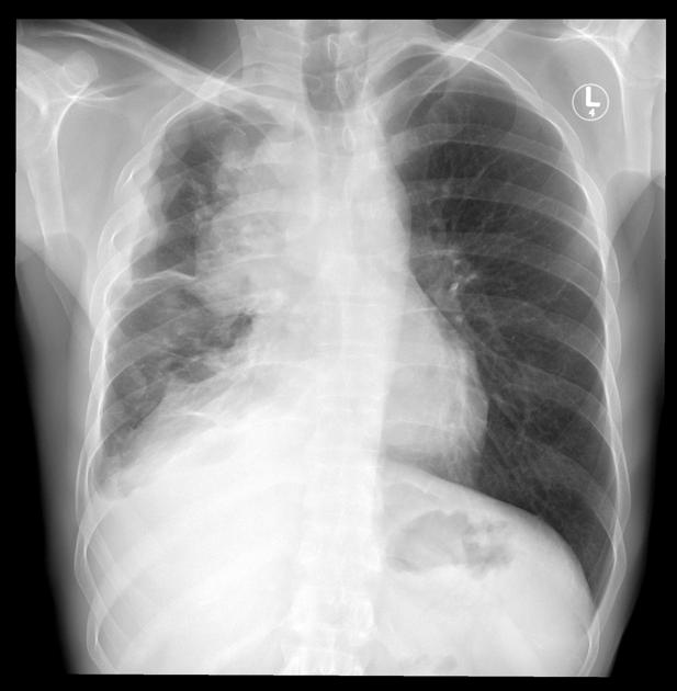
Anterior mediastinal mass. Right sided pleural effusion. Loss of volume of the right hemithorax, ? partial lung collapse. Hyperinflated left lung is clear.
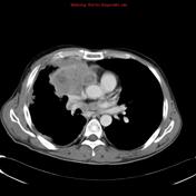

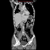

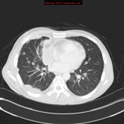

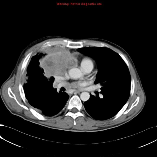
Bulky large anterior mediastinal mass with necrotic foci. Malignant nodular pleural thickening encasing the right hemithorax also involving the pericardium, with a small pericardial effusion. No convincing transgression of the right hemidiaphragm by tumor.
Multiple right-sided pulmonary nodules consistent with metastases.
Compensatory hyperinflation of the left lung. No left lung nodules or pleural disease.
No concerning findings beneath the diaphragm; prominent focal hepatic fat adjacent to the fissure for the ligamentum venosum.
Conclusion:
Invasive mediastinal mass with direct involvement of the pleura, pericardium, and multiple pulmonary metastases. No distant metastases of the contralateral lung or extrathoracic structures.
Case Discussion
Invasive thymoma with pleural metastasis mimicking pleural mesothelioma.


 Unable to process the form. Check for errors and try again.