
Cases
By sharing our collective experience through interesting and classic patient cases, we can make a real difference in how people are imaged and diagnosed. Each case belongs to a contributing member and all cases are reviewed by our dedicated editors to ensure they reach quality standards and abide by privacy guidelines. Cases can public or unlisted and then be viewed directly or added to articles, playlists or multiple choice questions. Find out more about cases.


58,341 results found
Case
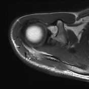
SLAP tear type 2
Published
23 Apr 2024
80% complete
MRI
Case
Spontaneous pneumothorax

Published
23 Apr 2024
94% complete
X-ray
Case
Intrapelvic cup migration

Published
23 Apr 2024
91% complete
X-ray
Case
Cerebral small vessel disease (illustration)

Published
23 Apr 2024
44% complete
Diagram
Case
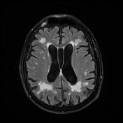
Cerebral microangiopathy
Published
23 Apr 2024
95% complete
MRI
Case
Brain death (SPECT)

Published
23 Apr 2024
94% complete
Nuclear medicine
Case
Post cholecystectomy biloma

Published
23 Apr 2024
92% complete
CT
Case
Shiny corner sign - ankylosing spondylitis

Published
23 Apr 2024
77% complete
MRI
Case
Longitudinal split tear of peroneus brevis

Published
23 Apr 2024
92% complete
MRI
Case
Cystitis cystica

Published
23 Apr 2024
92% complete
CT
Case
Multiple bone infarcts

Published
23 Apr 2024
73% complete
MRI
Case
Sinding-Larsen-Johansson disease

Published
23 Apr 2024
79% complete
X-ray
Case
Colocolonic intussusception

Published
23 Apr 2024
92% complete
Ultrasound
CT
Case
Androgen insensitivity syndrome

Published
22 Apr 2024
92% complete
MRI
Case
Occult femoral neck fracture

Published
22 Apr 2024
70% complete
X-ray
CT
MRI
Case
Anterior knee dislocation and popliteal artery injury

Published
22 Apr 2024
86% complete
X-ray
DSA (angiography)
CT
Case
Lymphoid interstitial pneumonia

Published
22 Apr 2024
80% complete
CT
Case
Ureteric jet

Published
22 Apr 2024
88% complete
Ultrasound
Case
Slit ventricle syndrome

Published
22 Apr 2024
80% complete
MRI
Case
Hematocolpos

Published
22 Apr 2024
77% complete
CT
ADVERTISEMENT: Supporters see fewer/no ads


 Unable to process the form. Check for errors and try again.