braın
Playlist contributed by
HMB *
Play
Share
Playlist
Full screen playlist
Playlist with hidden diagnosis
Full screen playlist with hidden diagnosis
Playlist information
Playlist created:
29 Oct 2023 by
HMB *
Last edited:
11 Nov 2023
Number of cases:
69
Number of slides:
0
rID:
178016
Systems:
Cardiac
,
Central Nervous System
,
Chest
,
Head & Neck
,
Musculoskeletal
,
Oncology
,
Paediatrics
,
Vascular
Visibility:
public
Show case titles
Case 1
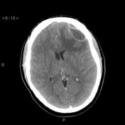
CT head bone window axial skull base - labeling questions
Case 2
Teaching head CT with annotated scrollable images
Case 3
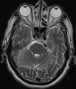
Trigeminal neuralgia due to vertebrobasilar dolichoectasia
Case 4
CT angiogram head axial - labeling questions
Case 5
CT angiogram head axial - labeling questions
Case 6
CT angiogram head sagittal - labeling questions
Case 7
Normal sinus CT (annotated)
Case 8
Normal petrous temporal bone axial CT - with labels
Case 9
Dural venous sinuses (illustration)
Case 10
Normal petrous temporal bone axial CT - with labels
Case 11
Skull landmarks - annotated images
Case 12
Midbrain anatomy
Case 13
Jugular foramen (illustration)
Case 14
Fibrous dysplasia - skull base
Case 15
Normal cranial nerves
Case 16
CT facial bones/orbits sagittal - labeling questions
Case 17
Subcortical U-fibers and juxtacortical lesions (illustration)
Case 18
Corpus callosum (annotated)
Case 19
Parieto-occipital region (diagram)
Case 20
Internal cerebral vein (annotated DSA)
Case 21
AC-PC line (diagram)
Case 22
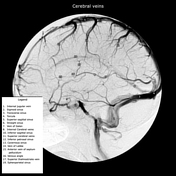
Cerebral veins (annotated DSA)
Case 23
Left temporal bone (illustration)
Case 24
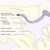
Pineal region anatomy (illustration)
Case 25
Middle cerebral artery branches
Case 26
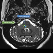
Abducens and facial cranial nerves and nuclei
Case 27
Layers of the scalp and meninges (illustrations)
Case 28
Cerebellar tonsillar position (illustration)
Case 29
Cavernous sinus (diagram)
Case 30
Posterior fossa vascular territories (illustration)
Case 31
Cerebral veins (diagram)
Case 32
Anatomy: sulci of the brain
Case 33
Basal vein of Rosenthal (annotated image)
Case 34
Frontal horn, intercaudate and inner table ratios (diagram)
Case 35
CT head axial - labeling questions
Case 36
CT chest arterial phase sagittal - labeling questions
Case 37
CT chest arterial phase coronal - labeling questions
Case 38
CT chest arterial phase axial - labeling questions
Case 39
CT neck with annotated scrollable images
Case 40
Teaching head CT with annotated scrollable images
Case 41
Pituitary MRI - normal study
Case 42
Pituitary MRI - normal study
Case 43
Normal neonatal CT head
Case 44
CT head bone window axial calvarium - labeling questions
Case 45
CT angiogram head coronal - labeling questions
Case 46
CT facial bones/orbits coronal - labeling questions
Case 47
Normal temporal bone CT with annotated images
Case 48
CT angiogram head axial - labeling questions
Case 49
Fazekas scale for white matter lesions
Case 50
Normal MRI hip
Case 51
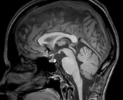
MRI head sagittal T1 - labeling questions
Case 52
Sinugenic epidural empyema
Case 53
Pituitary MRI - normal study
Case 54
External ear anatomy: annotated CT
Case 55
Neuroblastoma with CNS metastases
Case 56
Periosteal reactions (diagram)
Case 57
Illustration of a "dural tail" sign
Case 58
Skull landmarks - annotated images
Case 59
Epstein-Barr virus encephalitis
Case 60
Corpus callosum agenesis (illustrations)
Case 61
Cochlear implant
Case 62
Sagittal midline of the brain - normal anatomy
Case 63
Normal cranial nerves
Case 64
Oculomotor nerve (normal)
Case 65
Kernohan phenomenon
Case 66
Chordoma - thumbing the pons sign (illustration)
Case 67
Horizontal gaze palsy with progressive scoliosis (HGPPS)
Case 68
Tuberous sclerosis
Case 69
Tuberous sclerosis with enhancing radial band