
Search results for “abdominal ct ”


206 results found
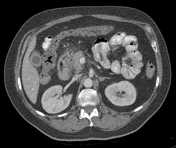
Case
Phantom abdominal CT

Published
14 Jan 2020
21% complete
CT
Photo
Article
Pancreas
The pancreas (plural: pancreata) is an unpaired, mostly retroperitoneal organ that has endocrine and exocrine functions, with a role in glucose metabolism and digestion.
Gross anatomy
Location
The pancreas is located at approximately the L1-L2 vertebral level in the anterior pararenal space o...
Article
Sonographic halo sign (disambiguation)
Sonographic halo sign can be useful in a number of situations:
hypoechoic halo sign (also known as target or bull's eye sign) in liver metastases: used in hepatobiliary imaging, is a concerning feature for malignant lesion if the lesion is a hyperechoic liver lesion 1,2
ultrasound halo in angi...
Article
Ascites
Ascites (hydroperitoneum is a rare synonym) is defined as an abnormal amount of intraperitoneal fluid.
Terminology
Ascites (plural is the same word) tends to be reserved for relatively sizable amounts of peritoneal fluid. The amount has not been defined formally. It is noted physiologically, h...
Article
Abdominal tuberculosis
Abdominal tuberculosis can manifest in almost every abdominopelvic organ:
gastrointestinal tuberculosis
esophageal tuberculosis
gastric tuberculosis
duodenal tuberculosis
jejunal and ileal tuberculosis
ileocecal tuberculosis
colorectal tuberculosis
tuberculous pe...
Article
Abdominal compartment syndrome
Abdominal compartment syndrome is a disease defined by the presence of new end-organ dysfunction secondary to elevated intra-abdominal pressure. Radiological diagnosis is difficult and usually suggested when a collection of imaging findings are present in the appropriate clinical setting or if t...
Article
CT abdomen-pelvis (protocol)
The CT abdomen-pelvis protocol serves as an outline for an examination of the whole abdomen including the pelvis. It is one of the most common CT protocols for any clinical questions related to the abdomen and/or in routine and emergencies. It forms also an integral part of trauma and oncologic ...
Article
Increased splenic density
Increased splenic density can be due to a number of processes. The density may be due to calcification (most common) or other compounds (iron, Thorotrast), and can be seen (often incidentally) on abdominal radiographs and CT. On CT the usual splenic attenuation is 35-55 HU or ~10 HU 6 lower than...
Article
Sarcoidosis (abdominal manifestations)
Sarcoidosis is a systemic inflammatory disease of unknown origin characterized by the formation of non-caseating granulomas. Virtually any organ system may be involved. Although less common than pulmonary and mediastinal disease, abdominal sarcoidosis can mimic more common infectious or neoplast...
Question
Question 2470
A 50-year-old woman presents with upper abdominal pain and undergoes CT of the abdomen and pelvis. There is a cystic lesion in the pancreas with no septations or enhancement, and a visible connection to the normal caliber pancreatic duct. What is the most likely diagnosis?
Article
Bile duct injury
Bile duct injuries are a potentially serious surgical problem associated with high morbidity, mortality, and prolonged hospitalization 1,2. These injuries typically occur infrequently as a complication of technically difficult laparoscopic cholecystectomy procedures or in the setting of hepatobi...
Article
Porcelain gallbladder
Porcelain gallbladder refers to extensive calcium encrustation of the gallbladder wall. The term has been used to emphasize the blue discolouration and brittle consistency of the gallbladder wall at surgery but is often an incidental finding on multiple different imaging modalities.
Clinical p...
Article
Acute abdominal pain
Acute abdominal pain is a common acute presentation in clinical practice. It encompasses a very broad range of possible etiologies and diagnoses, and imaging is routinely employed as the primary investigative tool in its modern management.
Terminology
A subgroup of patients with acute abdomina...
Case
Retained gallstone

Published
07 Jun 2017
95% complete
MRI
CT
Annotated image
Case
Bleeding duodenal ulcer and adenomyomatosis of gallbladder
Published
20 Jul 2018
95% complete
Ultrasound
CT
DSA (angiography)
Case
Pseudocirrhosis

Published
07 Jul 2017
95% complete
CT
Case
Ruptured renal hydatid cyst

Published
16 Nov 2020
92% complete
CT
Case
Mercedes-Benz sign of gallstones

Published
29 Jul 2016
89% complete
CT
Case
Hepatic pseudocirrhosis

Published
09 Jul 2017
75% complete
CT
Article
Pancreas transplant
A pancreas transplant is a major surgical procedure in which a donor pancreas is transplanted into a recipient. The donor pancreas is typically cadaveric, but may rarely be a segment from a living donor 1. The transplant is meant to establish normoglycemia in patients with diabetes mellitus, typ...


 Unable to process the form. Check for errors and try again.