


3,754 results found
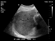
Case
Hepatic hydatid cyst - stage CE4

Published
15 May 2024
75% complete
Ultrasound
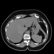
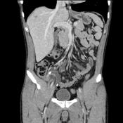
Case
Post cholecystectomy clips

Published
16 May 2024
95% complete
CT
Case
Undifferentiated embryonal sarcoma of the liver

Published
31 Dec 2017
80% complete
Ultrasound
CT
Case
Hepatic hydatid cyst compressing the left portal vein

Published
20 May 2019
80% complete
CT
Ultrasound
Article
Hypervascular liver lesions
Hypervascular liver lesions are findings that enhance more or similarly to the background hepatic parenchyma in the late arterial phase, on contrast-enhanced CT or MRI.
Differential diagnosis
Non-neoplastic
focal nodular hyperplasia (FNH)
bright arterial phase enhancement except central scar...
Article
Sinusoidal obstruction syndrome
Sinusoidal obstruction syndrome (SOS), previously known as hepatic veno-occlusive disease (VOD), is a condition arising from occlusion of hepatic venules.
Clinical presentation
right upper quadrant pain
painful hepatomegaly
ascites
abnormal liver function tests
Pathology
Toxic injury to l...
Article
Hepatic leiomyosarcoma
Hepatic leiomyosarcomas are rare primary malignant tumors derived from smooth muscle cells in the liver.
Epidemiology
Hepatic leiomyosarcoma is rare 1. An equal sex distribution and a broad age range (5 months-66Y) has been reported. Some have suggested an associated with AIDS 2.
Pathology
T...
Article
Pancreas transplant
A pancreas transplant is a major surgical procedure in which a donor pancreas is transplanted into a recipient. The donor pancreas is typically cadaveric, but may rarely be a segment from a living donor 1. The transplant is meant to establish normoglycemia in patients with diabetes mellitus, typ...
Case
Cirrhosis
Published
13 May 2024
72% complete
Ultrasound
Case
Focal nodular hyperplasia

Published
23 May 2023
74% complete
MRI
Ultrasound
Case
Adenomyomatosis of the gallbladder - fundal

Published
09 Jun 2023
77% complete
MRI
Ultrasound
Article
Hepatic hemangioma
Hepatic hemangiomas or hepatic venous malformations are the most common benign vascular liver lesions. They are frequently diagnosed as an incidental finding on imaging, and most patients are asymptomatic. From a radiologic perspective, it is important to differentiate hemangiomas from hepatic m...
Case
Cardiac failure and the deer horn sign

Published
06 Nov 2023
94% complete
Ultrasound
Case
Biphasic portal vein Doppler trace

Published
02 Jan 2018
69% complete
Ultrasound
Article
Beaver tail liver
Beaver tail liver, also known as a sliver of liver, is a variant of hepatic morphology where an elongated left liver lobe extends laterally to contact and often surround the anterior aspect of the spleen 2.
Beaver tail liver is more common in females. The parenchyma is normal and therefore has ...
Case
Elephant (Rorschach radiology)
Published
05 May 2024
65% complete
CT
Article
Whole-body CT (protocol)
CT polytrauma/multitrauma, also called trauma CT, whole body CT (WBCT) or panscan, is an increasingly used investigation in patients with multiple injuries sustained after significant trauma.
The majority of the evidence regarding whole-body CT is, understandably, retrospective. There is some e...
Article
Liver trauma
The liver is one of the most frequently damaged organs in blunt trauma, and liver trauma is associated with a significant mortality rate.
Epidemiology
In blunt abdominal trauma, the liver is injured ~5% (range 1-10%) of the time 1,3.
Clinical presentation
Patients can present with right uppe...
Case
Acute appendicitis
Published
19 Jan 2014
86% complete
CT
Case
Bouveret syndrome

Published
22 Feb 2019
95% complete
X-ray
Photo
Annotated image
CT


 Unable to process the form. Check for errors and try again.