

21,802 results found
Case
Stieda fracture

Published
19 Jan 2022
91% complete
X-ray
Case
Acromioclavicular joint ganglion and long head of biceps brachii dislocation

Published
06 Feb 2011
63% complete
Ultrasound
Case
Gamekeeper thumb
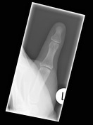
Published
24 Jan 2018
91% complete
X-ray
Case
Parameniscal cyst

Published
09 Jul 2022
68% complete
X-ray
MRI
Case
Metacarpal fracture

Published
18 Jan 2022
94% complete
X-ray
Case
Right pubic rami fractures

Published
16 Oct 2014
91% complete
X-ray
Article
Lateral talocalcaneal angle
The lateral talocalcaneal angle is one of the angles that can be measured for the assessment of pes planus and pes cavus and assessment of hindfoot deformity.
Measurement
The lateral talocalcaneal angle is drawn on a weight-bearing lateral foot radiograph. There are two ways that it has been d...
Case
Calcaneofibular ligament bony avulsion

Published
07 May 2023
91% complete
X-ray
Ultrasound
Case
Anterior shoulder dislocation

Published
09 Jan 2017
88% complete
X-ray
Case
Hoffa fracture of the lateral femoral condyle

Published
10 Mar 2020
89% complete
MRI
Case
Left upper lobe pneumonia and pectus excavatum

Published
25 May 2013
79% complete
X-ray
Case
Rolando fracture

Published
19 Nov 2023
94% complete
X-ray
Article
Chordoma
Chordomas are uncommon malignant tumors of the axial skeleton that account for 1% of intracranial tumors and 4% of all primary bone tumors.
They originate from embryonic remnants of the primitive notochord (earliest fetal axial skeleton, extending from the Rathke's pouch to the tip of the cocc...
Article
Internal oblique muscle
The internal oblique muscle is one of the muscles that form the anterior abdominal wall. Inferiorly, it contributes towards the formation of the inguinal ligament.
Summary
origin: originates along the whole length of the lumbar fascia, from the anterior two-thirds of the intermediate line of t...
Case
Superior lumbar hernia

Published
06 May 2013
83% complete
CT
Article
Transversus abdominis muscle
The transversus abdominis muscle, named according to the direction of its muscle fibers, is one of the flat muscles that form the anterior abdominal wall. It is deep to the internal oblique muscle and ends in the anterior aponeurosis, which ultimately blends with the linea alba.
Summary
origi...
Case
Supracondylar fracture

Published
27 Mar 2022
94% complete
X-ray
Article
External oblique muscle
The external oblique muscle is one of the muscles that forms the anterior abdominal wall. Its free inferior border forms the inguinal ligament, and its aponeurotic part contributes to the anterior wall of the inguinal canal.
Summary
origin: outer surface of the shaft of the lower eight ribs 3...
Article
Congenital diaphragmatic hernia
Congenital diaphragmatic herniation (CDH) accounts for a small proportion of all diaphragmatic herniae. However, it is one of the most common non-cardiac fetal intrathoracic anomalies.
Epidemiology
Congenital diaphragmatic hernias are seen in 1 of every 2000-4000 live births. 84% are left-side...
Case
Dorsal defect of the patella with cartilage involvement and bone marrow edema

Published
27 Apr 2022
74% complete
MRI


 Unable to process the form. Check for errors and try again.