


21,687 results found
Article
Posterior tibial line
The posterior tibial line is drawn along the posterior aspect of the distal tibial shaft on a lateral ankle x-ray and can be used to assess the sagittal alignment of the talus when comparing side-to-side and/or calculate the posterior tibial line-talar ratio 1,2.
Case
Stener lesion

Published
07 Jun 2016
69% complete
Ultrasound
Case
Ramp lesion

Published
04 Aug 2022
74% complete
MRI
Case
Atlanto-occipital assimilation with basilar invagination

Published
12 Aug 2019
80% complete
MRI
Case
S1 transforaminal epidural steroid injection (fluoroscopic guided)

Published
14 May 2021
63% complete
Fluoroscopy
Article
Chronic ACL deficiency
Chronic ACL deficiency is a long term adverse outcome that can result from an untreated ACL injury. Patients often experience an unstable knee. They often result in or are associated with meniscal injury (medial more than lateral) and chondral damage 1-4.
See also
mucoid degeneration of ACL
Case
Multiple myeloma

Published
04 Mar 2014
75% complete
CT
Article
Periosteal chondrosarcoma
Periosteal chondrosarcomas, previously also known as juxta-cortical chondrosarcomas, are cartilagineous or chondroid matrix-generating neoplasms originating in close association with the periosteum from the bony surface 1-3.
Terminology
The term ‘juxta-cortical chondrosarcoma’ is no longer rec...
Article
Osteonecrosis of the femoral head
Osteonecrosis of the femoral head, previously known as avascular necrosis (AVN) of the hip, is the most common site for osteonecrosis, presumably due to a combination of precarious blood supply and high loading when standing.
Idiopathic osteonecrosis of the femoral head epiphysis in children (...
Case
Ulnar fracture and radial head dislocation

Published
16 May 2016
94% complete
X-ray
Case
Foreign body in heel

Published
28 Oct 2012
66% complete
Ultrasound
Case
Posterior dislocation of sternoclavicular joint

Published
27 Feb 2020
89% complete
CT
X-ray
Case
Flexor carpi ulnaris - calcific tendinitis

Published
11 Feb 2022
74% complete
Photo
Ultrasound
Case
Spondylolysis - L4

Published
19 Sep 2014
80% complete
CT
Case
Os supranaviculare

Published
05 Nov 2023
75% complete
X-ray
Case
Paget disease of the skull

Published
07 May 2008
75% complete
CT
Case
Tumoral calcinosis

Published
03 Sep 2020
75% complete
X-ray
Case
Proximal focal femoral deficiency - bilateral class A

Published
25 Jan 2022
80% complete
CT
Case
Normal left wrist radiograph

Published
15 Apr 2022
69% complete
X-ray
Case
Paget disease - skull
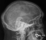
Published
03 Nov 2009
60% complete
X-ray


 Unable to process the form. Check for errors and try again.