


9,356 results found
Case
Traumatic aortic injury

Published
03 Oct 2016
90% complete
CT
X-ray
DSA (angiography)
Article
Extrapleural fat sign
The extrapleural fat sign is an imaging feature which can be seen on CT under certain circumstances. It occurs from the inward displacement of extrapleural fat by an extrapleural fluid collection, extrapleural hematoma or extrapleural mass. The presence of the extrapleural fat sign is indicative...
Case
Pulmonary septic emboli

Published
12 May 2021
92% complete
CT
X-ray
Article
Medical devices in the thorax
Medical devices in the thorax are regularly observed by radiologists when reviewing radiographs and CT scans.
Extrathoracic devices
tubing, clamps, syringes, scissors, lying on or under the patient
rubber sheets, foam mattresses, clothing, hair braids, nipple piercings, etc., may also be visi...
Case
Pneumomediastinum

Published
17 Apr 2022
94% complete
X-ray
Case
Bifid left fourth rib and hypoplastic right fifth rib

Published
11 Nov 2023
91% complete
X-ray
Case
Misplaced nasogastric tube

Published
17 Apr 2022
94% complete
X-ray
Article
Subcutaneous emphysema
Subcutaneous emphysema (also known commonly, although less correctly, as surgical emphysema), strictly speaking, refers to gas in the subcutaneous tissues. But the term is generally used to describe any soft tissue emphysema of the body wall or limbs since the gas often dissects into the deeper ...
Case
Diaphragm rupture

Published
07 Dec 2022
89% complete
CT
X-ray
Case
Tuberculosis

Published
27 Apr 2023
92% complete
CT
Ultrasound
Case
Potassium chloride tablet in trachea

Published
07 May 2008
95% complete
CT
Article
Bronchogenic cyst
Bronchogenic cysts are congenital malformations of the bronchial tree (a type of bronchopulmonary foregut malformation). They can present as a mediastinal mass that may enlarge and cause local compression. It is also considered the commonest of foregut duplication cysts.
Epidemiology
Bronchoge...
Case
Pneumothorax

Published
05 Mar 2024
88% complete
Ultrasound
Case
Scleroderma - thoracic and gastrointestinal manifestations

Published
12 Jan 2018
95% complete
CT
Case
Incidental lung nodules
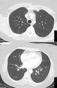
Published
29 Jun 2012
53% complete
CT
Case
Usual interstitial pneumonia

Published
22 Apr 2022
86% complete
CT
Article
Garland triad
Garland triad, also known as the 1-2-3 sign or pawnbroker's sign, is a lymph node enlargement pattern on chest radiographs which has been described in sarcoidosis:
right paratracheal nodes
right hilar nodes
left hilar nodes
Hilar lymphadenopathy is symmetrical and usually massive...
Case
Pulmonary embolism

Published
06 Nov 2014
92% complete
CT
Article
Right paratracheal lymphadenopathy
Right paratracheal lymphadenopathy represents pathological involvement of any of the lymph nodes in the right upper (2R) and/or lower (4R) paratracheal nodal groups 1. These nodes are often also enlarged but this is not always the case.
The commonest causes are sarcoidosis, tuberculosis and lun...
Case
Intercostal nerves (Gray's illustrations)

Published
27 Jan 2022
32% complete
Diagram


 Unable to process the form. Check for errors and try again.