
Cleft epiphysis
Updates to Article Attributes
Cleft epiphysis is a normal epiphyseal variant. It can be either unilateral or bilateral . The The most common site is basal epiphysisthe prepiphysis of the first proximal phalanx of great toe the foot.
Radiographic features
Plain filmradiograph
ShowsPlain radiographs will demonstrate a lucent defect in the epiphyseal region epiphysis. ItThe borders of the lucency are variable and may showbe sharp as well asor irregular borders . CleftThe cleft remains till the fusion of growth plate.
Clinical importance Differential diagnosis
HasA cleft epiphysis has to be differentiated from fracture. Generally fractures demonstrate signs of healing if x raythe radiograph is repeated after 2-3 weeks after injury. Recognition of this entity is important to avoid overtreatmentover treatment and unnecessary surgical intervention.
-<p><strong>Cleft epiphysis</strong> is a normal epiphyseal variant. It can be either unilateral or bilateral . The most common site is basal epiphysis of proximal phalanx of great toe .</p><h4>Radiographic features</h4><h5>Plain film</h5><p>Shows lucent defect in the epiphyseal region . It may show sharp as well as irregular borders . Cleft remains till the fusion of growth plate .</p><h4>Clinical importance </h4><p>Has to be differentiated from fracture. Generally fractures demonstrate signs of healing if x ray is repeated after 2-3 weeks. Recognition of this entity is important to avoid overtreatment and unnecessary surgical intervention.</p>- +<p><strong>Cleft epiphysis</strong> is a normal epiphyseal variant. It can be either unilateral or bilateral The most common site is the prepiphysis of the first proximal phalanx of the foot.</p><h4>Radiographic features</h4><h5>Plain radiograph</h5><p>Plain radiographs will demonstrate a lucent defect in the epiphysis. The borders of the lucency are variable and may be sharp or irregular. The cleft remains till the fusion of growth plate.</p><h4>Differential diagnosis</h4><p>A cleft epiphysis has to be differentiated from fracture. Generally fractures demonstrate signs of healing if the radiograph is repeated 2-3 weeks after injury. Recognition of this entity is important to avoid over treatment and unnecessary surgical intervention.</p>
References changed:
- 3. Berquist TH. Imaging of the Foot and Ankle. Lippincott Williams & Wilkins. (2012) ISBN:1451147996. <a href="http://books.google.com/books?vid=ISBN1451147996">Read it at Google Books</a> - <a href="http://www.amazon.com/gp/product/1451147996">Find it at Amazon</a><span class="auto"></span>
Tags changed:
- variant
Sections changed:
- Anatomy
Image 1 X-ray (Frontal) ( create )

Image 2 X-ray (Frontal) ( update )
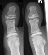
Image 3 X-ray (Coned frontal) ( create )

Image 4 X-ray (Frontal) ( create )


 Unable to process the form. Check for errors and try again.