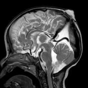
Atretic parietal cephalocele
Updates to Primarylink Attributes
Updates to Synonym Attributes
Updates to Article Attributes
Body
was changed:
Atretic parietal cephalocele (APC) also known as atertic cephalocele refers to small subscalp lesion which consists of dura, fibrous tissue, and dysplastic brain tissue.
Epidemiology
Common presentation in infants and young children.
Pathology
It is thought to represent involuted true cephalocele (meningocele or encephalocele) connected to dura matter via a fibrous stalk.
Clinical presentation
Palpable midline parietal soft tissue mass.
Associations
Increased incidence of intracranial anomalies.
Radiographic features
- subgaleal soft tissue mass with intracranial extension via sharply demarcated calvarial defect (cranium bifidum)
- CSF tract and vertical falcine vein point to the subcutaneous scalp mass
- vertically oriented primitive falcine vein
- fibrous stalk connecting the cephalocele
- focal fenestration of superior sagittal sinus at the atretic parietal cephalocele
- prominence of superior cerebellar cistern and suprapineal recess
- superior peaking of the posterior tentorium
- spinning top configuration of the tentorial incisura
Differential diagnosis
- sinus pericranii
- dermoid or epidermoid cyst
- cephalohematoma
- sebaceous cyst
- vascular lesions (hemangioma)
Prognosis
Prognosis of atertic cephalocels is generally good.
see also
-<p><strong>Atretic parietal c</strong><strong>ephalocele</strong> (<strong>APC</strong>) also known as <strong>atertic cephalocele</strong> refers to small subscalp lesion which consists of dura, fibrous tissue, and dysplastic brain tissue.</p><h4>Epidemiology</h4><p>Common presentation in infants and young children. </p><h4>Pathology</h4><p>It is thought to represent involuted true cephalocele (meningocele or encephalocele) connected to dura matter via a fibrous stalk. </p><h4>Clinical presentation</h4><h5><span style="font-size:13px; font-weight:normal; line-height:20.7999992370605px">Palpable midline parietal soft tissue mass.</span></h5><h5>Associations</h5><p>Increased incidence of intracranial anomalies.</p><h4>Radiographic features</h4><ul>- +<p><strong>Atretic parietal c</strong><strong>ephalocele</strong> (<strong>APC</strong>) also known as <strong>atertic </strong><a href="/articles/cephalocoele">cephalocele</a> refers to small subscalp lesion which consists of dura, fibrous tissue, and dysplastic brain tissue.</p><h4>Epidemiology</h4><p>Common presentation in infants and young children. </p><h4>Pathology</h4><p>It is thought to represent involuted true cephalocele (meningocele or encephalocele) connected to dura matter via a fibrous stalk.<strong> </strong></p><h4>Clinical presentation</h4><p>Palpable midline parietal soft tissue mass.</p><h5>Associations</h5><p>Increased incidence of intracranial anomalies.</p><h4>Radiographic features</h4><ul>
-</ul><h4>Prognosis</h4><p>Prognosis of atertic cephalocels is good.</p>- +</ul><h4>Prognosis</h4><p>Prognosis of atertic cephalocels is generally good.</p><h5>see also</h5><p><a href="/articles/cephalocoele">Cephalocoele</a></p>
References changed:
- 1. Osborn AG, Salzman KL, Barkovich AJ. Diagnostic Imaging: Brain. Lippincott Williams & Wilkins. (2009) ISBN:1931884722. <a href="http://books.google.com/books?vid=ISBN1931884722">Read it at Google Books</a> - <a href="http://www.amazon.com/gp/product/1931884722">Find it at Amazon</a><span class="ref_v3"></span>
- 2. Wong SL, Law HL, Tan S. Atretic cephalocele - an uncommon cause of cystic scalp mass. Malays J Med Sci. 2012;17 (3): 61-3. <a href="http://www.ncbi.nlm.nih.gov/pmc/articles/PMC3216175">Free text at pubmed</a> - <a href="http://www.ncbi.nlm.nih.gov/pubmed/22135551">Pubmed citation</a><span class="auto"></span>
- 3. Muralidharan CG, Aggarwal R, Singh D. Atretic parietal encephalocoele - An unusual diagnosis. Med J Armed Forces India. 2013;69 (1): 83-5. <a href="http://dx.doi.org/10.1016/j.mjafi.2012.02.005">doi:10.1016/j.mjafi.2012.02.005</a> - <a href="http://www.ncbi.nlm.nih.gov/pmc/articles/PMC3862940">Free text at pubmed</a> - <a href="http://www.ncbi.nlm.nih.gov/pubmed/24532943">Pubmed citation</a><span class="auto"></span>
Systems changed:
- Central Nervous System
Images Changes:
Image 1 MRI (T2) ( create )