
Breast tissue markers
Updates to Article Attributes
Body
was changed:
workBreast tissue markers are a common finding in progress - DSbreast radiology. These are typically inserted following percutaneous biopsy, either under ultrasound or sterotactic guidance. They can be invaluable in identifying known benign areas or shrinking/treated malignant lesions on follow up imaging.
A number of various makers are in use including ribbon, coil and wing markers with easily differentiated appearances on mammograms. Some new markers are available with superior detection on ultrasound.
Markers can also be used to target intervention (localisation wire targets) or with clips inserted peri-operatively to guide subsequent radiotherapy treatments.
-<p>work in progress - DS</p>- +<p><strong>Breast tissue markers</strong> are a common finding in breast radiology. These are typically inserted following percutaneous biopsy, either under ultrasound or sterotactic guidance. They can be invaluable in identifying known benign areas or shrinking/treated malignant lesions on follow up imaging.</p><p>A number of various makers are in use including ribbon, coil and wing markers with easily differentiated appearances on mammograms. Some new markers are available with superior detection on ultrasound.</p><p>Markers can also be used to target intervention (localisation wire targets) or with clips inserted peri-operatively to guide subsequent radiotherapy treatments.</p>
References changed:
- 1. Thomassin-Naggara I, Lalonde L, David J, Darai E, Uzan S, Trop I. A plea for the biopsy marker: how, why and why not clipping after breast biopsy?. (2012) Breast cancer research and treatment. 132 (3): 881-93. <a href="https://doi.org/10.1007/s10549-011-1847-x">doi:10.1007/s10549-011-1847-x</a> - <a href="https://www.ncbi.nlm.nih.gov/pubmed/22042370">Pubmed</a> <span class="ref_v4"></span>
Tags changed:
- stub
Systems changed:
- Breast
Images Changes:
Image 1 Annotated image (Left CC zoomed) ( create )
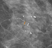
Image 2 Ultrasound (Left lower inner quadrant) ( create )

Image 3 Annotated image (CC zoomed) ( create )

Image 4 Annotated image (CC zoomed) ( create )

Image 5 Annotated image (CC zoomed) ( create )

Caption
was changed:
Case 45: coil marker and wire
Position
was set to
.
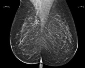
Image 6 Mammography (MLO) ( create )
Caption
was changed:
Case 56: post-operative surgical clips
Position
was set to
.


 Unable to process the form. Check for errors and try again.