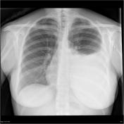
Chest x-ray review: breathing
Updates to Article Attributes
-
thisThis is abasic article
forfor medical students and other non-radiologists
Chest x-ray review is is a key competency for medical students, junior doctors and other allied health professionals.
Using A, B, C, D, E
-
A:airwayis a helpful and systematic method for chest x-ray review -
where B
: breathing (i.e.refers to breathing and the assessment of the lungs) -
C: circulation (i.e. cardiomediastinum) -
D: disability (i.e. fracturesanddislocations) -
E: everything else
pleural spaces.Breathing
There are a large number of pathological process that can result in abnormality in the lungs. This is usually highlighted as a change in the appearance of normally aeration. Broadly speaking, this may be because of increased density density (white areas) or increased lucency lucency (black areas).
While the mediastinum isn't central, you should still be able to compare the lungs, ensuring that the apices, upper, middle and lower zones are of similar density and volume.
When looking and comparing the lung parenchyma, check for any focal areas of increased density.
After checking the lung parenchyma, check around the lungs. Start laterally, from the apex down to the costophrenic angle. After checking the costophrenic angle and making sure they are symmetrical, ensure you can trace the hemidiaphragms to the spine. If the hemidiaphragms are flattened, there may be hyperinflation. Finally Finally, check the cardiac borders up to the hilar structures.
Checklist
- both lungs are expanded and similar in volume
- apices, upper, middle and lower zones are symmetrical
- normal lateral margins
- normal CPAs
- normal hemidiaphragms
- normal cardiac borders
- normal lung behind the heart
Pathology
The pathology that affects the lung parenchyma is wide ranging.
hyperinflation-
airspace shadowingconsolidation/pneumoniapulmonary oedema
-
collapselung collapse-
lobar collapseright upper lobe collapseright middle lobe collapseright lower lobe collapseleft upper lobe collapseleft lower lobe collapse
segmental collapsesubsegmental collapselung fibrosis
-
lung lesionssolitary pulmonary lesionmultiple lung lesionscavitating lung lesion
-
pleural collectionpneumonectomypleural effusionhaemothoraxempyema
-<ul><li>this is a <em>basic article</em> for medical students and non-radiologists</li></ul><p><strong>Chest x-ray review</strong> is a key competency for medical students, junior doctors and other allied health professionals.</p><h4>A, B, C, D, E</h4><ul>-<li>-<strong>A</strong>: <a title="Chest x-ray A (basic)" href="/articles/chest-x-ray-a-basic">airway</a>-</li>-<li>-<strong>B</strong>: breathing (i.e. the lungs)</li>-<li>-<strong>C</strong>: circulation (i.e. cardiomediastinum)</li>-<li>-<strong>D</strong>: disability (i.e. fractures and dislocations)</li>-<li>-<strong>E</strong>: everything else</li>-</ul><h5>Breathing</h5><p>There are a large number of pathological process that can result in abnormality in the lungs. This is usually highlighted as a change in the appearance of normally aeration. Broadly speaking, this may be because of increased density (white areas) or increased lucency (black areas).</p><p>While the mediastinum isn't central, you should still be able to compare the lungs, ensuring that the apices, upper, middle and lower zones are of similar density and volume.</p><p>When looking and comparing the lung parenchyma, check for any focal areas of increased density.</p><p>After checking the lung parenchyma, check around the lungs. Start laterally, from the apex down to the costophrenic angle. After checking the costophrenic angle and making sure they are symmetrical, ensure you can trace the hemidiaphragms to the spine. If the hemidiaphragms are flattened, there may be hyperinflation. Finally, check the cardiac borders up to the hilar structures.</p><h6>Checklist</h6><ul>- +<h6>This is a basic article for medical students and other non-radiologists</h6><p><strong>Chest x-ray review</strong> is a key competency for medical students, junior doctors and other allied health professionals. Using A, B, C, D, E is a helpful and systematic method for <a href="/articles/chest-x-ray-basic-an-approach">chest x-ray review</a> where B refers to breathing and the assessment of the lungs and pleural spaces.</p><h4>Breathing</h4><p>There are a large number of pathological process that can result in abnormality in the lungs. This is usually highlighted as a change in the appearance of normally aeration. Broadly speaking, this may be because of increased density (white areas) or increased lucency (black areas).</p><p>While the mediastinum isn't central, you should still be able to compare the lungs, ensuring that the apices, upper, middle and lower zones are of similar density and volume.</p><p>When looking and comparing the lung parenchyma, check for any focal areas of increased density.</p><p>After checking the lung parenchyma, check around the lungs. Start laterally, from the apex down to the costophrenic angle. After checking the costophrenic angle and making sure they are symmetrical, ensure you can trace the hemidiaphragms to the spine. If the hemidiaphragms are flattened, there may be hyperinflation. Finally, check the cardiac borders up to the hilar structures.</p><h5>Checklist</h5><ul>
-</ul><h6>Pathology</h6><p>The pathology that affects the lung parenchyma is wide ranging.</p><ul>-<li>hyperinflation</li>-<li>airspace shadowing<ul>-<li>consolidation/pneumonia</li>-<li>pulmonary oedema</li>-</ul>-</li>-<li>collapse<ul>-<li>lung collapse</li>-<li>-<a href="/articles/lobar-collapse-basic">lobar collapse</a><ul>-<li>right upper lobe collapse</li>-<li>right middle lobe collapse</li>-<li>right lower lobe collapse</li>-<li>left upper lobe collapse</li>-<li>left lower lobe collapse</li>-</ul>-</li>-<li><a href="/articles/segmental-collapse-basic">segmental collapse</a></li>-<li>subsegmental collapse</li>-<li>lung fibrosis</li>-</ul>-</li>-<li>lung lesions<ul>-<li>solitary pulmonary lesion</li>-<li>multiple lung lesions</li>-<li>cavitating lung lesion</li>-</ul>-</li>-<li>pleural collection<ul>-<li>pneumonectomy</li>-<li>pleural effusion</li>-<li>haemothorax</li>-<li>empyema</li>-</ul>-</li>
Systems changed:
- Chest
Image 1 X-ray (PA) ( create )

Image 2 X-ray (Frontal) ( create )

Image 3 X-ray (Frontal) ( create )

Image 4 X-ray (Frontal) ( create )

Image 5 X-ray (Frontal) ( create )

Image 6 X-ray ( create )

Image 7 X-ray (Frontal) ( create )

Image 8 Annotated image ( create )

Image 9 Annotated image ( create )

Image 10 Annotated image ( create )

Image 11 Annotated image ( create )

Image 12 Annotated image ( create )

Image 13 Annotated image ( create )

Image 14 Annotated image ( create )

Image 15 Annotated image ( create )


 Unable to process the form. Check for errors and try again.