Dural ectasia
Updates to Article Attributes
Dural ectasia refers to ballooning or widening of the dural sac which can result in posterior vertebral scalloping and is associated with herniation of nerve root sleeves.
Clinical presentation
Patients with dural ectasia may present with low back pain or radicular pain in the buttocks or legs. Pain may be accompanied by leg weakness or urinary incontinence. Other associations include spondylolisthesis, scoliosis, vertebral erosions, and vertebral fractures. There is also an increased incidence of anterior sacral meningocoeles which may present as an abdominal mass.
Associations
- Marfan syndrome: dural ectasia has been observed in 60-90% of patients; in these patients, the dilatation of the dural sac is almost always in the lumbar region
- neurofibromatosis type 1
- Ehlers-Danlos syndrome
- ankylosing spondylitis
- osteogenesis imperfecta
- trauma
- post surgery
- tumours
- scoliosis
Radiographic features
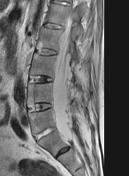
Dural ectasia is dilationdescribes widening of the dural sac or spinal nerve root sleeves, usually associated with bony erosions of the posterior vertebral body 4. AnteroposteriorOne study suggests that the anteroposterior diameter of the thecal sac at the S1 level should be greater than that of the thecal sac at the L4 level in order to describe dural ectasia ref required4.
Plain radiograph
Posterior vertebral scalloping may be an indirect indicator 1-2,4. However, this is not specific, as it is seen in a significant percentage of the normal population and is also associated with several other conditions.
MRI
Increase in the APanteroposterior diameter of the dural sac, usually in the lumbar region4.
Differential diagnosis
-</ul><h4>Radiographic features</h4><p>Dural ectasia is dilation of the dural sac. Anteroposterior diameter of the thecal sac at the S1 level greater than that of the thecal sac at the L4 level <sup>ref required</sup>.</p><h5>Plain radiograph</h5><p>Posterior <a href="/articles/vertebral-scalloping">vertebral scalloping</a> may be an indirect indicator <sup>1-2</sup>. However, this is not specific, as it is seen in a significant percentage of the normal population and is also associated with several other conditions.</p><h5>MRI</h5><p>Increase in the AP diameter of the dural sac, usually in the lumbar region.</p><h4>Differential diagnosis</h4><ul><li><a href="/articles/tarlov-cyst">Tarlov cyst</a></li></ul>- +</ul><h4>Radiographic features</h4><p>Dural ectasia describes widening of the dural sac or spinal nerve root sleeves, usually associated with bony erosions of the posterior vertebral body <sup>4</sup>. One study suggests that the anteroposterior diameter of the thecal sac at the S1 level should be greater than that of the thecal sac at the L4 level in order to describe dural ectasia <sup>4</sup>.</p><h5>Plain radiograph</h5><p>Posterior <a href="/articles/vertebral-scalloping">vertebral scalloping</a> may be an indirect indicator <sup>1-2,4</sup>. However, this is not specific, as it is seen in a significant percentage of the normal population and is also associated with several other conditions.</p><h5>MRI</h5><p>Increase in the anteroposterior diameter of the dural sac, usually in the lumbar region <sup>4</sup>.</p><h4>Differential diagnosis</h4><ul><li><a href="/articles/tarlov-cyst">Tarlov cyst</a></li></ul>
References changed:
- 4. Lundby R, Rand-Hendriksen S, Hald JK, Lilleås FG, Pripp AH, Skaar S, Paus B, Geiran O, Smith H-J. Dural Ectasia in Marfan Syndrome: A Case Control Study. American Journal of Neuroradiology. 30 (8): 1534. <a href="https://doi.org/10.3174/ajnr.A1620">doi:10.3174/ajnr.A1620</a> - <a href="https://www.ncbi.nlm.nih.gov/pubmed/19461064">Pubmed</a> <span class="ref_v4"></span>
Image 3 MRI (T2) ( update )

Image 6 MRI (T2) ( create )