
Intramuscular lipomas are deep-seated lipomas located within a muscle.
On this page:
Terminology
Intramuscular lipomas share the term ‘infiltrating lipoma’ with intermuscular lipomas.
Epidemiology
Intramuscular lipomas account for about 1% of all lipomas and occur in all age groups with most occurring between the 5th to 7th decades of life. There seems to be a female predominance 1.
Clinical presentation
Patients might present with unspecific swelling of the tumor or be found incidentally. Rare presentations are pain or paresthesia due to nerve entrapment 1,2.
Pathology
Intramuscular lipomas consist of mature adipocytes identical to normal adult fatty tissue 1, the exact etiology remains unclear.
Subtypes
infiltrative
well-defined or circumscribed
Location
They can occur at any anatomical site but are believed to occur mainly in the large muscles of the limbs and the trunk, an exact anatomical distribution, however, has not been established. Intramuscular lipomas are rare in the hand and feet and very rare in the oral cavity 1,3.
Macroscopic appearance
Intramuscular lipomas are usually yellowish and located within muscle. They may show a capsule at the margin that can show interdigitations and muscle fibers passing through 1.
Microscopic appearance
Mature adipocytes irregularly infiltrate and replace muscle fibers and bundles in the infiltrative type 1. There is no fatty infiltration of adjacent muscle typically observed in the well-defined or well-circumscribed type.
Radiographic features
Plain radiograph
An intramuscular lipoma can appear as a lucency, especially if large and can show radioopaque streaks.
Ultrasound
Appears as a hyperechoic, well-defined mass with fine internal echoes or striated appearance due to interdigitations of muscular tissue.
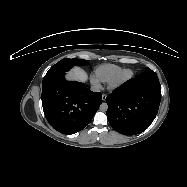
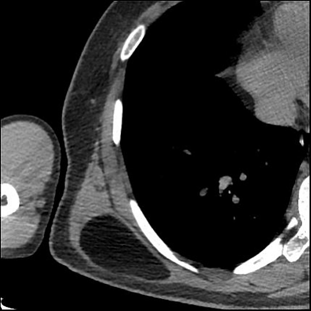
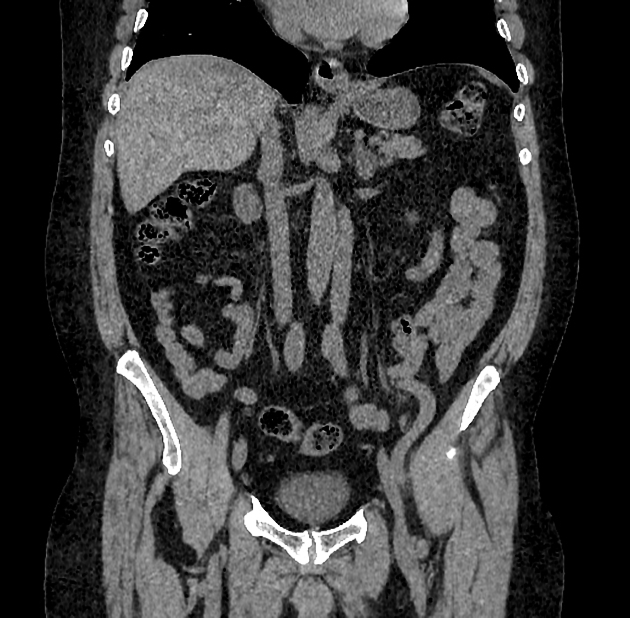
CT
Intramuscular lipomas usually show the following features 1:
hypodense soft tissue mass within the musculature
typically with Hounsfield measurements in the negative range (fat density)
can show a striated appearance
can show thick intramuscular septae and interdigitations
usually, oval or fusiform shape but can vary
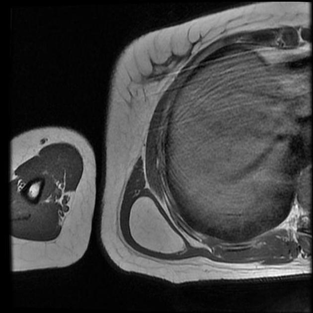
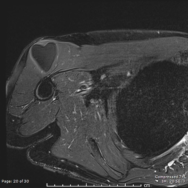
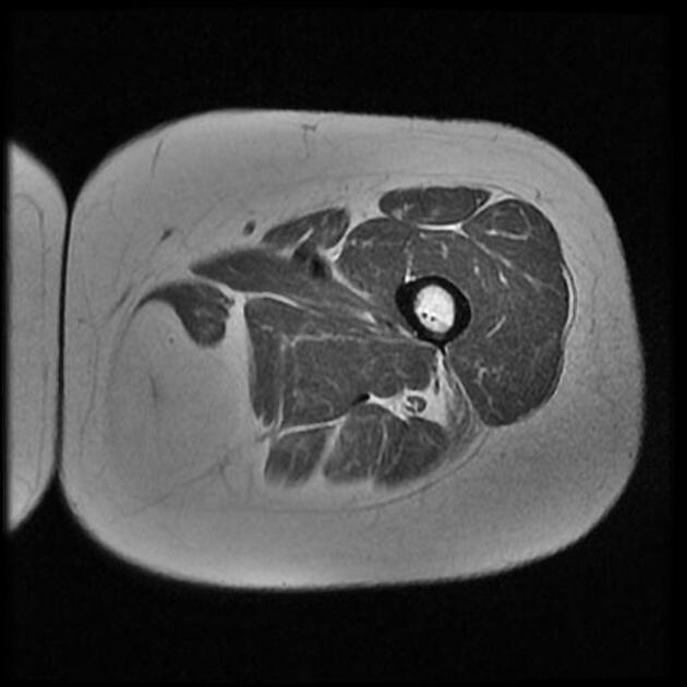
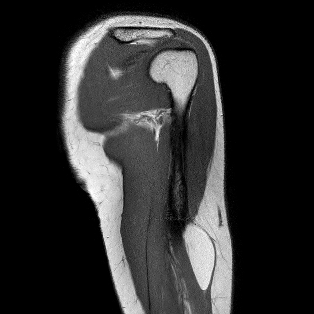
MRI
Usually shows either a fat-containing mass within a muscle, which is isointense to subcutaneous fat in all sequences sometimes with septae. Septae should not enhance avidly however or show nodules 2. Interlocked or intermingled muscular tissue and fibers can be observed in the infiltrative type 1. If a capsule is present and found outside the intramuscular lipoma, there should not be muscular fibers in the main mass 1.
Treatment and prognosis
Treatment depends on tumor size location and symptoms and includes watchful waiting or wide resection.
Differential diagnosis
intermuscular lipoma: located between muscles
well-differentiated liposarcoma (dumb-bell shaped, should be suspected if septae markedly enhance)


 Unable to process the form. Check for errors and try again.