
Koerner septum
Citation, DOI, disclosures and article data
Citation:
Priamo F, Weerakkody Y, Bell D, et al. Koerner septum. Reference article, Radiopaedia.org (Accessed on 22 Mar 2025) https://doi.org/10.53347/rID-54470
rID:
54470
Article created:
10 Jul 2017,
Francesco Priamo
Disclosures:
At the time the article was created Francesco Priamo had no recorded disclosures.
View Francesco Priamo's current disclosures
Last revised:
Disclosures:
At the time the article was last revised Yuranga Weerakkody had no financial relationships to ineligible companies to disclose.
View Yuranga Weerakkody's current disclosures
Revisions:
13 times, by
5 contributors -
see full revision history and disclosures
Systems:
Sections:
Synonyms:
- Koerner septum
- Korner's septum
- Koerner septa
- Koerner's septum
- Koerner's septa
- Korner septum
- Körner's septum
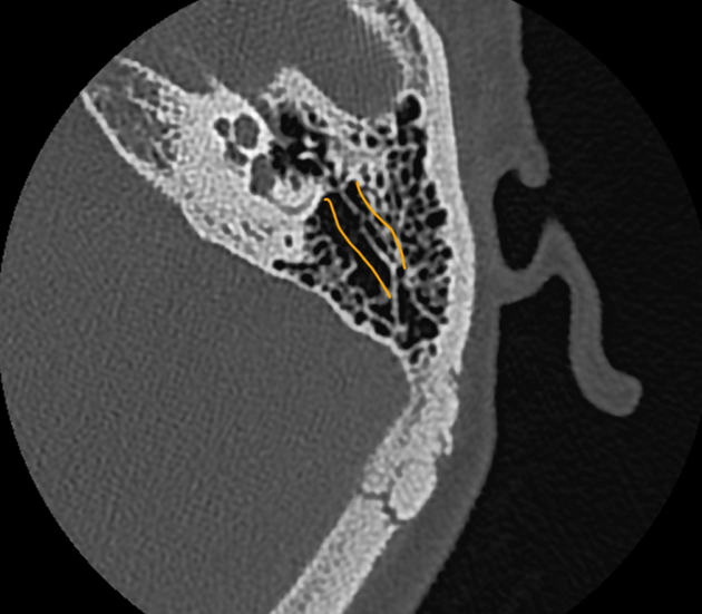
The Koerner septum, also known as the petrosquamous lamina or septum, is a thin bridge of bone which divides the petrous and squamous portion of the mastoid air cells at the level of the mastoid antrum. It represents the continuation and persistence of the petrosquamous suture 2.
It is commonly eroded by middle ear cholesteatomas.
History and etymology
It is named after Otto Körner (1858-1935), a German otorhinolaryngologist, described the septum petrosquamosum in 1926 3.
References
- 1. Gaurano JL, Joharjy IA. Middle ear cholesteatoma: characteristic CT findings in 64 patients. Annals of Saudi medicine. 24 (6): 442-7. Pubmed
- 2. Wojciechowski T, Skadorwa T, Drożdż A, Ciszek B, Szopiński K. The radioanatomical assessment of the Körner's septum. (2019) Surgical and radiologic anatomy : SRA. 41 (6): 669-673. doi:10.1007/s00276-018-2149-3 - Pubmed
- 3. Körner O. Das Septum petrosquamosum (mastoideum) und seine klinische Bedeutung. Fortschr Hals Nasen Ohrenheilkd. 1926;17:137–140.
Incoming Links
Related articles: Anatomy: Head and neck
- skeleton of the head and neck
-
cranial vault
- scalp (mnemonic)
- fontanelle
-
sutures
- calvarial
- facial
- frontozygomatic suture
- frontomaxillary suture
- frontolacrimal suture
- frontonasal suture
- temporozygomatic suture
- zygomaticomaxillary suture
- parietotemporal suture (parietomastoid suture)
- occipitotemporal suture (occipitomastoid suture)
- sphenofrontal suture
- sphenozygomatic suture
- spheno-occipital suture (not a true suture)
- lacrimomaxillary suture
- nasomaxillary suture
- internasal suture
- basal/internal
- skull landmarks
- frontal bone
- temporal bone
- parietal bone
- occipital bone
- skull base (foramina)
-
facial bones
- midline single bones
- paired bilateral bones
- cervical spine
- hyoid bone
- laryngeal cartilages
-
cranial vault
- muscles of the head and neck
- muscles of the tongue (mnemonic)
- muscles of mastication
-
facial muscles
- epicranius muscle
- circumorbital and palpebral muscles
- nasal muscles
-
buccolabial muscles
- elevators, retractors and evertors of the upper lip
- levator labii superioris alaeque nasalis muscle
- levator labii superioris muscle
- zygomaticus major muscle
- zygomaticus minor muscle
- levator anguli oris muscle
- malaris muscle
- risorius muscle
- depressors, retractors and evertors of the lower lip
- depressor labii inferioris muscle
- depressor anguli oris muscle
- mentalis muscle
- compound sphincter
-
orbicularis oris muscle
- incisivus labii superioris muscle
- incisivus labii inferioris muscle
-
orbicularis oris muscle
- muscle of mastication
- modiolus
- elevators, retractors and evertors of the upper lip
- muscles of the middle ear
- orbital muscles
- muscles of the soft palate
- pharyngeal muscles
- suprahyoid muscles
- infrahyoid muscles
- intrinsic muscles of the larynx
- muscles of the neck
- platysma muscle
- longus colli muscle
- longus capitis muscle
- scalenus anterior muscle
- scalenus medius muscle
- scalenus posterior muscle
- scalenus pleuralis muscle
- sternocleidomastoid muscle
-
suboccipital muscles
- rectus capitis posterior major muscle
- rectus capitis posterior minor muscle
- obliquus capitis superior muscle
- obliquus capitis inferior muscle
- accessory muscles of the neck
- deep cervical fascia
-
deep spaces of the neck
- anterior cervical space
- buccal space
- carotid space
- danger space
- deep cervical fascia
- infratemporal fossa
- masticator space
- parapharyngeal space
- stylomandibular tunnel
- parotid space
- pharyngeal (superficial) mucosal space
- perivertebral space
- posterior cervical space
- pterygopalatine fossa
- retropharyngeal space
- suprasternal space (of Burns)
- visceral space
- surgical triangles of the neck
- orbit
- ear
- paranasal sinuses
- upper respiratory tract
- viscera of the neck
- blood supply of the head and neck
-
arterial supply
-
common carotid artery
- carotid body
- carotid bifurcation
- subclavian artery
- variants
-
common carotid artery
- venous drainage
-
arterial supply
- innervation of the head and neck
-
cranial nerves
- olfactory nerve (CN I)
- optic nerve (CN II)
- oculomotor nerve (CN III)
- trochlear nerve (CN IV)
-
trigeminal nerve (CN V) (mnemonic)
- trigeminal ganglion
- ophthalmic division
- maxillary division
- mandibular division
- abducens nerve (CN VI)
- facial nerve (CN VII)
-
vestibulocochlear nerve (CN VIII)
- vestibular ganglion (Scarpa's ganglion)
- glossopharyngeal nerve (CN IX)
- vagus nerve (CN X)
- (spinal) accessory nerve (CN XI)
- hypoglossal nerve (CN XII)
- parasympathetic ganglia of the head and neck
- cervical sympathetic ganglia
- greater occipital nerve
- third occipital nerve
-
cervical plexus
- muscular branches
- longus capitis
- longus colli
- scalenes
- geniohyoid
- thyrohyoid
-
ansa cervicalis
- omohyoid (superior and inferior bellies separately)
- sternothyroid
- sternohyoid
- phrenic nerve
- contribution to the accessory nerve (CN XI)
- cutaneous branches
- muscular branches
- brachial plexus
- pharyngeal plexus
-
cranial nerves
- lymphatic drainage of the head and neck
- embryological development of the head and neck


 Unable to process the form. Check for errors and try again.