Morton neuroma
Updates to Article Attributes
Morton neuromas are focal areas of symptomatic perineural fibrosis around a plantar digital nerve of the foot. The abnormality is non-neoplastic and does not represent a true neuroma. It may more correctly be known as Morton’s metatarsalgia. The condition is thought to be due to chronic entrapment of the nerve by the intermetatarsal ligament.
Epidemiology
It most often occurs in middle-aged individuals and is many times more common in women than men. Approximately 30% of asymptomatic middle-aged persons have the radiologic / pathologic findings of a Morton’s neuroma. Symptomatic lesions tend to be slightly larger (mean 5.3mm versus 4.1mm in one large series 1). Lesions greater than 5 mm are very likely to be symptomatic. 10% of lesions are bilateral.
Clinical presentation
Patient's typically present with forefoot pain which radiates from mid foot to toes 5. Symptoms are often progressive and worsened by activity.
Location
The 3rd web-space (between 3rd and 4thmetatarsal heads) is the most commonly affected site. The 2nd web-space less commonly involved while the remaining web-spaces are rarely involved.
Pathology
It is characterised by neural degeneration with epineural and endoneural vascular hyalinization, and perineural fibrosis around an intermetatarsal nerve 2.
Radiographic features
Ultrasound
Typically seen as a round to ovoid, well-defined, hypo-echoic lesion in the intermetatarsal space adjacent are proximal to the metatarsal head 4. A small proportion can have mixed echotexture 5.
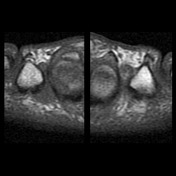
MRI
Dumbell / ovoid shaped lesion at a similar position to that described on ultrasound.
- T1: typically low to iso signal 1-2
- T2: typically low signal but can sometimes be intermediate in signal
- T1 C+ (Gd): tends to show intense enhancement
Treatment and prognosis
Ultrasound-guided interdigital injection of steroid and local anaesthetic has been shown to have a relatively good success rate.9
Surgical excision can also be performed, also with a relatively good success rate (~ 80% 6).
EtymologyHistory and etymology
It was initially described by Thomas Morton in 1876 4
Differential diagnosis
MR Imaging differential considerations include
- changes secondary to a plantar plate tear - disruption 7-8
-</ul><h4>Treatment and prognosis</h4><p>Ultrasound-guided interdigital injection of steroid and local anaesthetic has been shown to have a relatively good success rate.<sup>9</sup> </p><p>Surgical excision can also be performed, also with a relatively good success rate (~ 80% <sup>6</sup>).</p><h4>Etymology</h4><p>It was initially described by <strong>Thomas Morton</strong> in 1876 <sup>4</sup></p><h4>Differential diagnosis</h4><p>MR Imaging differential considerations include</p><ul><li>changes secondary to a <a href="/articles/plantar-plate-tear">plantar plate tear</a> - disruption <sup>7-8</sup>- +</ul><h4>Treatment and prognosis</h4><p>Ultrasound-guided interdigital injection of steroid and local anaesthetic has been shown to have a relatively good success rate.<sup>9</sup> </p><p>Surgical excision can also be performed, also with a relatively good success rate (~ 80% <sup>6</sup>).</p><h4>History and etymology</h4><p>It was initially described by <strong>Thomas Morton</strong> in 1876 <sup>4</sup></p><h4>Differential diagnosis</h4><p>MR Imaging differential considerations include</p><ul><li>changes secondary to a <a href="/articles/plantar-plate-tear">plantar plate tear</a> - disruption <sup>7-8</sup>
Image 1 Annotated image (Coronal T1) ( update )

Image 2 MRI (T1) ( update )