
Mosaic attenuation pattern in lung
Updates to Article Attributes
Body
was changed:
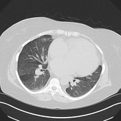
Mosaic attenuationis the description given to the appearance at CT where there is a patchwork of regions of differing attenuation. It is a non-specific finding, which may be seen in any of the following:
- obstructive small airways disease: low attenuation regions are abnormal and reflect decreased perfusion of the poorly ventilated regions, e.g. bronchiectasis, cystic fibrosis, constrictive bronchiolitis
- occlusive vascular disease (can be termed a mosaic perfusion pattern in this setting 7): low attenuation regions are abnormal and reflect relative oligaemia, e.g. chronic pulmonary embolism
- parenchymal disease: high attenuation regions are abnormal and represent ground-glass opacity
Differentiating the cause
This term "mosaic attenuation" is non-specific, and best used where the differentiation between mosaic perfusion (or mosaic oliagemia) and ground-glass opacity cannot be confidently made.
However, ascertaining the underlying cause for mosaic attenuation is often possible on the basis of clinical information, combined with the assessment of other lung features on HRCT2,5:
- peripheral vessels: if vessels in hypoattenuated regions of the lung are smaller than in the other regions, the pattern is due to mosaic perfusion (i.e. airways or vascular disease rather than ground-glass)
- central vessels: pulmonary hypertension, reflected as dilatation of the central pulmonary arteries, suggests a vascular cause
- small airways: the presence of abnormally dilated or thick walled airways in the relatively lucent lung confirms underlying airway disease, see small airways disease
- parenchymal changes: ground glass opacity is the likely cause for mosaic attenuation if other features of infiltrative disease are present, such as reticular opacities (i.e. crazy paving pattern) or nodules
- air trapping: refers to regions of lung which following expiration do not show the normal increase in attenuation, or show little change in cross-sectional area5 (i.e. this is an expiratory HRCT finding); the presence of air trapping suggests airway disease
-<p><strong>Mosaic attenuation </strong>is the description given to the appearance at CT where there is a patchwork of regions of differing attenuation. It is a non-specific finding, which may be seen in any of the following:</p><ul>- +<p><strong>Mosaic attenuation </strong>is the description given to the appearance at CT where there is a patchwork of regions of differing attenuation. It is a non-specific finding, which may be seen in any of the following:</p><ul>
-<strong>obstructive small airways disease:</strong> low attenuation regions are abnormal and reflect decreased perfusion of the poorly ventilated regions, e.g. <a href="/articles/bronchiectasis">bronchiectasis</a>, <a href="/articles/pulmonary-manifestations-of-cystic-fibrosis">cystic fibrosis</a>, <a href="/articles/constrictive-bronchiolitis">constrictive bronchiolitis </a>- +<strong>obstructive small airways disease:</strong> low attenuation regions are abnormal and reflect decreased perfusion of the poorly ventilated regions, e.g. <a href="/articles/bronchiectasis">bronchiectasis</a>, <a href="/articles/cystic-fibrosis-pulmonary-manifestations-1">cystic fibrosis</a>, <a href="/articles/constrictive-bronchiolitis">constrictive bronchiolitis </a>
-<strong>parenchymal disease:</strong> high attenuation regions are abnormal and represent <a href="/articles/ground-glass-opacification">ground-glass opacity</a>- +<strong>parenchymal disease:</strong> high attenuation regions are abnormal and represent <a href="/articles/ground-glass-opacification-1">ground-glass opacity</a>
-</ul><h4>Differentiating the cause</h4><p>This term "mosaic attenuation" is non-specific, and best used where the differentiation between mosaic perfusion (or mosaic oliagemia) and ground-glass opacity cannot be confidently made.</p><p>However, ascertaining the underlying cause for mosaic attenuation is often possible on the basis of clinical information, combined with the assessment of other lung features on HRCT <sup>2,5</sup>:</p><ol>- +</ul><h4>Differentiating the cause</h4><p>This term "mosaic attenuation" is non-specific, and best used where the differentiation between mosaic perfusion (or mosaic oliagemia) and ground-glass opacity cannot be confidently made.</p><p>However, ascertaining the underlying cause for mosaic attenuation is often possible on the basis of clinical information, combined with the assessment of other lung features on HRCT <sup>2,5</sup>:</p><ol>
-<strong><a href="/articles/air-trapping">air trapping</a>:</strong> refers to regions of lung which following expiration do not show the normal increase in attenuation, or show little change in cross-sectional area <sup>5</sup> (i.e. this is an <a href="/articles/expiratory-hrct-1">expiratory HRCT</a> finding); the presence of air trapping suggests airway disease</li>- +<strong><a href="/articles/air-trapping">air trapping</a>:</strong> refers to regions of lung which following expiration do not show the normal increase in attenuation, or show little change in cross-sectional area <sup>5</sup> (i.e. this is an <a href="/articles/expiratory-hrct-1">expiratory HRCT</a> finding); the presence of air trapping suggests airway disease</li>
Images Changes:
Image 5 CT (lung window) ( create )


 Unable to process the form. Check for errors and try again.