Pneumopericardium
Updates to Article Attributes
Pneumopericardium represents air within the pericardium, thus surrounding the heart.
Underlying causes include:
- thoracic surgery
/ pericardial/pericardial fluid drainage - penetrating trauma
- blunt trauma (rare)
- infectious peticarditis with gas-producing organisms
- fistula
- between the pericardium and an adjacent air-containing organ (i.e. stomach or esophagus)
Radiographic features
Plain radiograph and CT
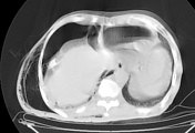
On both chest radiographs and CT, appearances are characteristic, the heart being partially or completely surrounded by air, with the pericardium sharply outlined by air density on either side.
Complications
Differential diagnosis
A pneumopericardium can usually be distinguished from pneumomediastinum, since air in the pericardial sac should not rise above the anatomic limits of the pericardial reflection on the proximal great vascular pedicle. Also on radiographs obtained with the patient in the decubitus position, air in the pericardial sac will shift immediately, while air in the mediastinum will not shift in a short interval between films.
Occasionally, it may not be possible to distinguish pneumopenicardium from pneumomediastinum on plain film.
-<li>thoracic surgery / pericardial fluid drainage</li>- +<li>thoracic surgery/pericardial fluid drainage</li>
Image 3 CT (lung window) ( update )