Radial neck fracture
Updates to Article Attributes
Radial neck fractures are, together with the radial head fractures, relatively common injuries, especially in adults, although they can be occult on radiographs.
Pathology
Radial neck fractures are almost always the result of a fall onto an outstretched hand. Force applied along the radius results in impaction of the radial head against the capitellum. The result is often a radial head or neck fracture. The neck fractures may be complete or incomplete.
Radiographic features
Plain radiograph
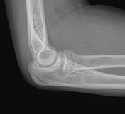
The elbow is typically radiographed in AP and lateral projections, although an oblique view is very frequently also obtained to better visualise the radial head (see elbow radiography).
The images generally show the loss of the mild concave curvature of the anterior cortex of the base of the radial head creating an abrupt offset between the radial head and neck 1.
An elbow joint effusion will almost alwaysis usually present, however, it could be presentabsent on plain x-ray in some cases as part of the radial neck is extra-articular.
Treatment and prognosis
Radial neck fractures are usually not displaced. As a result, they are usually treated with immobilisation in cast. In the rare situation where there is a transverse fracture of the neck and displacement of the proximal fracture fragment, operative intervention may be required.
-<p><strong>Radial neck fractures</strong> are, together with the <a href="/articles/radial-head-fractures">radial head fractures</a>, relatively common injuries, especially in adults, although they can be occult on radiographs. </p><h4>Pathology</h4><p>Radial neck fractures are almost always the result of a fall onto an outstretched hand. Force applied along the radius results in impaction of the radial head against the capitellum. The result is often a radial head or neck fracture. The neck fractures may be complete or incomplete.</p><h4>Radiographic features</h4><h5>Plain radiograph</h5><p>The elbow is typically radiographed in AP and lateral projections, although an oblique view is very frequently also obtained to better visualise the radial head (see <a href="/articles/elbow-series">elbow radiography</a>).</p><p>The images generally show the loss of the mild concave curvature of the anterior cortex of the base of the radial head creating an abrupt offset between the radial head and neck <sup>1</sup>. An <a href="/articles/elbow-joint-effusion">elbow joint effusion</a> will almost always be present. </p><h4>Treatment and prognosis</h4><p>Radial neck fractures are usually not displaced. As a result, they are usually treated with immobilisation in cast. In the rare situation where there is a transverse fracture of the neck and displacement of the proximal fracture fragment, operative intervention may be required.</p>- +<p><strong>Radial neck fractures</strong> are, together with the <a href="/articles/radial-head-fracture-2">radial head fractures</a>, relatively common injuries, especially in adults, although they can be occult on radiographs. </p><h4>Pathology</h4><p>Radial neck fractures are almost always the result of a fall onto an outstretched hand. Force applied along the radius results in impaction of the radial head against the capitellum. The result is often a radial head or neck fracture. The neck fractures may be complete or incomplete.</p><h4>Radiographic features</h4><h5>Plain radiograph</h5><p>The elbow is typically radiographed in AP and lateral projections, although an oblique view is very frequently also obtained to better visualise the radial head (see <a href="/articles/elbow-series">elbow radiography</a>).</p><p>The images generally show the loss of the mild concave curvature of the anterior cortex of the base of the radial head creating an abrupt offset between the radial head and neck <sup>1</sup>.</p><p>An <a href="/articles/elbow-joint-effusion">elbow joint effusion</a> is usually present, however, it could be absent on plain x-ray in some cases as part of the radial neck is extra-articular. </p><h4>Treatment and prognosis</h4><p>Radial neck fractures are usually not displaced. As a result, they are usually treated with immobilisation in cast. In the rare situation where there is a transverse fracture of the neck and displacement of the proximal fracture fragment, operative intervention may be required.</p>
Image 6 MRI (T1) ( destroy )
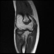
Image 6 MRI (PD fat sat) ( create )
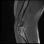
Image 7 X-ray (Lateral - zoomed) ( create )