Rhabdomyosarcomas (biliary tract)
Updates to Article Attributes
Rhabdomyosarcomas of the biliary tract are rare tumours, usually identified in children, with a very poor prognosis. TheyThey are usually grouped under botryoid rhabdomyosarcomas.
For a general discussion of this type of tumour, please refer to the article onrhabdomyosarcomas.
Epidemiology
Rhabdomyosarcomas are considered the most common biliary tract tumours in children 4. The median age of presentation is 3 years with a slight male preponderance.3.
Clinical presentation
The most common clinical features are jaundice and abdominal distention, with pain, vomiting, and fever being less frequent. Elevation in liver transaminases and bilirubin is often present.
Pathology
Location
Rhabdomyosarcomas of the biliary tract most commonly arises from the common bile duct 1, but it can arise from almost anywhere along the biliary tree including liver, intrahepatic and extrahepatic biliary ducts, gallbladder, or ampulla. It has also been reported to arise from hepatic and choledochal cysts.
Radiographic features
Ultrasonography
Ultrasound imaging will demonstrate intrahepatic biliary ductal dilatation and an intraductal mass. Cystic areas may be seen in larger tumours and may represent areas of necrosis3. Because of this, it can have a radiologic appearance similar to a choledochal cyst, especially if there is no local invasion.
CT
CT may show a heterogeneous or hypo-attenuatinghypoattenuating mass with biliary ductal dilatation. CT may be the best modality for surveillance of recurrence.
MRI
MRI has advantages over other modalities because of its ability to define the extent of disease and relationship to the hepatic vasculature.
Differential diagnosis
If the tumour is large and grows into the liver, it may be difficult to distinguish from other primary liver masses 3.
-<p><strong>Rhabdomyosarcomas of the biliary tract</strong> are rare tumours, usually identified in children, with a very poor prognosis. They are usually grouped under <a href="/articles/botryoid-rhabdomyosarcoma">botryoid rhabdomyosarcomas</a>.</p><p>For a general discussion of this type of tumour, please refer to the article on <a href="/articles/rhabdomyosarcoma">rhabdomyosarcomas</a>.</p><h4>Epidemiology</h4><p>Rhabdomyosarcomas are considered the most common biliary tract tumours in children <sup>4</sup>. The median age of presentation is 3 years with a slight male preponderance.<sup>3</sup></p><h4>Clinical presentation</h4><p>The most common clinical features are <a href="/articles/jaundice">jaundice</a> and abdominal distention, with pain, vomiting, and fever being less frequent. Elevation in liver transaminases and bilirubin is often present.</p><h4>Pathology</h4><h5>Location</h5><p>Rhabdomyosarcomas of the biliary tract most commonly arises from the common bile duct <sup>1</sup>, but it can arise from almost anywhere along the biliary tree including liver, intrahepatic and extrahepatic biliary ducts, gallbladder, or ampulla. It has also been reported to arise from hepatic and choledochal cysts.</p><h4>Radiographic features</h4><h5><strong>Ultrasonography</strong></h5><p>Ultrasound imaging will demonstrate intrahepatic biliary ductal dilatation and an intraductal mass . Cystic areas may be seen in larger tumours and may represent areas of necrosis<sup>3</sup>. Because of this, it can have a radiologic appearance similar to a <a href="/articles/choledochal-cyst">choledochal cyst</a>, especially if there is no local invasion.</p><h5><strong>CT</strong></h5><p>CT may show a heterogeneous or hypo-attenuating mass with biliary ductal dilatation. CT may be the best modality for surveillance of recurrence.</p><h5><strong>MRI</strong></h5><p>MRI has advantages over other modalities because of its ability to define the extent of disease and relationship to the hepatic vasculature.</p><h4>Differential diagnosis</h4><p>If the tumour is large and grows into the liver, it may be difficult to distinguish from other primary liver masses <sup>3</sup>.</p>- +<p><strong>Rhabdomyosarcomas of the biliary tract</strong> are rare tumours, usually identified in children, with a very poor prognosis. They are usually grouped under <a href="/articles/botryoid-rhabdomyosarcoma">botryoid rhabdomyosarcomas</a>.</p><p>For a general discussion of this type of tumour, please refer to the article on <a href="/articles/rhabdomyosarcoma">rhabdomyosarcomas</a>.</p><h4>Epidemiology</h4><p>Rhabdomyosarcomas are considered the most common biliary tract tumours in children <sup>4</sup>. The median age of presentation is 3 years with a slight male preponderance <sup>3</sup>.</p><h4>Clinical presentation</h4><p>The most common clinical features are <a href="/articles/jaundice">jaundice</a> and abdominal distention, with pain, vomiting, and fever being less frequent. Elevation in liver transaminases and bilirubin is often present.</p><h4>Pathology</h4><h5>Location</h5><p>Rhabdomyosarcomas of the biliary tract most commonly arises from the common bile duct <sup>1</sup>, but it can arise from almost anywhere along the biliary tree including liver, intrahepatic and extrahepatic biliary ducts, gallbladder, or ampulla. It has also been reported to arise from hepatic and choledochal cysts.</p><h4>Radiographic features</h4><h5>Ultrasound</h5><p>Ultrasound imaging will demonstrate intrahepatic biliary ductal dilatation and an intraductal mass. Cystic areas may be seen in larger tumours and may represent areas of necrosis<sup>3</sup>. Because of this, it can have a radiologic appearance similar to a <a href="/articles/choledochal-cyst">choledochal cyst</a>, especially if there is no local invasion.</p><h5>CT</h5><p>CT may show a heterogeneous or hypoattenuating mass with biliary ductal dilatation. CT may be the best modality for surveillance of recurrence.</p><h5>MRI</h5><p>MRI has advantages over other modalities because of its ability to define the extent of disease and relationship to the hepatic vasculature.</p><h4>Differential diagnosis</h4><p>If the tumour is large and grows into the liver, it may be difficult to distinguish from other primary liver masses <sup>3</sup>.</p>
Systems changed:
- Paediatrics
- Oncology
Image 1 Ultrasound (Transverse) ( create )
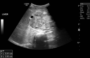
Image 2 CT (C+ portal venous phase) ( create )
