

Systematic review
Distal radial contour

Check the contour of the distal radius:
- AP
- the distal radial articular surface should cup the carpals
- the articular surface should get progressively more distal towards the radial styloid
- lateral
- the radial surface should be smooth
- there should be a palmar tilt to the articular surface
- if these features aren't present, think distal radial fracture
Carpal arcs

Check the carpal arcs:
- the articular surfaces of the proximal and distal carpal rows should form three smooth arcs
- trace these arcs on the AP film
- the spacing between all carpal bones should be 1-2 mm
- if the arc is broken or there is widening of a joint space, think carpal dislocation
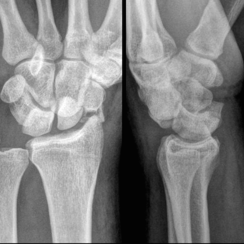
Carpal alignment (lateral)

Check lateral alignment:
- the distal radius, lunate and capitate should be in a straight line
- if the line has been disrupted, think:
- lunate dislocation: lunate is completely dislocated
- perilunate dislocation: lunate and capitate are dislocated
Carpometacarpal articulation

Check carpometacarpal joint space:
- a 1-2 mm joint space should be seen between the carpals and metacarpals
- if the joint space is narrowed, think carpometacarpal dislocation
One of the commonest misses in trauma films of the hand and wrist is dislocation of the 5th carpometacarpal joint which may cause significant morbidity if diagnosis is delayed.
Bone cortex

Check each bone in turn:
- pay particular attention to the distal radius, proximal carpal row (especially the scaphoid) and the bases of the metacarpals
Common pathology
Colles fracture

- the most common distal radial fracture in any adult group
- peak incidence in elderly women
- usually follow a fall onto an outstretched hand
- dorsal angulation of the distal fracture component
- important to determine if there is intra-article extension
- more: Colles fracture
Smith fracture
- account for less than 3% of forearm fractures
- more common in young males and elderly females
- fall onto flexed wrist or direct blow back of wrist
- classically, an extra-articular distal radius fracture with palmar angulation of distal fracture fragment
- also called the reverse Colles
- more: Smith fracture
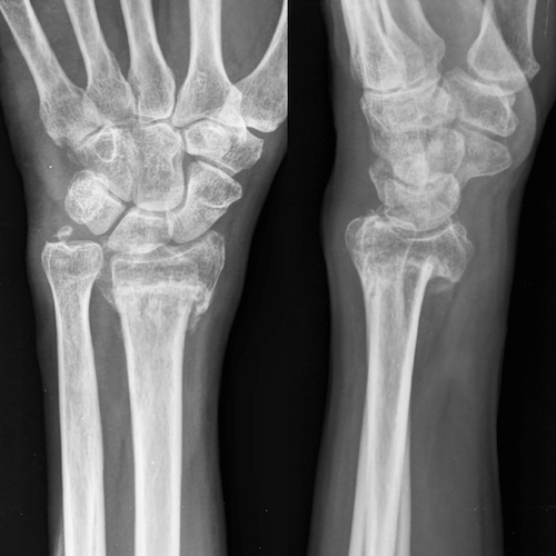
Scaphoid fracture
- 80% of all carpal bone fractures
- usually young adults
- fall on an outstretched hand
- 80% of fractures are through the waist of the scaphoid
- if suspected, perform additional scaphoid views
- may be radiographically occult - should be followed up if pain persists
- more: scaphoid fracture
Triquetral fracture

- second most common carpal bone fracture
- hyperextension or avulsive injury
- frequently seen as dorsal chip fractures on lateral views only
- more: triquetral fracture
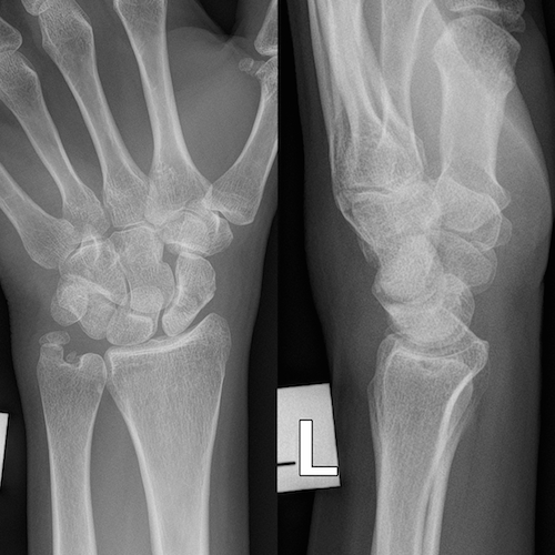
Perilunate dislocation
- typically occur in young adults
- following a fall on dorsiflexed wrist
- best detected on lateral view - the lunate articulates with distal radius but capitate does not sit in lunate ‘cup’
- 60% associated with scaphoid fracture
- more: perilunate dislocation
Don't miss...
Scapholunate dissociation

- injury to the scapholunate and radiolunate ligament results in scapholunate dissociation and significant instability
- the scapholunate space is widened (> 4 mm) - the Terry-Thomas sign
- more: scapholunate dissociation
Lunate dislocation

- much less common than perilunate dislocation
- commonly occur in young adults
- fall onto a dorsiflexed wrist
- more: lunate dislocation
Carpometacarpal dislocation
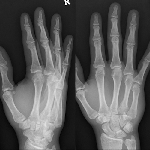
- rare but important injury to dominant hands of younger males
- younger male predominance
- often occur after a punch followed by a fall
- reduction of joint space on the AP
- best seen on an oblique study
- more: carpometacarpal dislocation


 Unable to process the form. Check for errors and try again.