Radiopaedia Blog

Practising radiology in the 21st Century might as well become a quite different affair than in the two previous centuries.

The ever changing world in which we live in has already heavily been influenced by  the world wide web (initially a pure scientific network) for quite a long time. With the more recent technology (smartphones, tablets) finding its way into working environment along with specialized applications ("Apps"), there already have been significant changes in the way radiologist work world wide, and more are very likely to be on their way.

The so far documented changes or changes probably to emerge include the way radiology residents and fellows:

  • learn and seek information
  • view imaging studies
  • communicate imaging findings to clinicians and patients

You yourself and the site you are currently viewing seem no exception to this evolutionary development.

The presently available resources already hold the opportunity to find up-to-date high-quality medical information at a split second and appear to be all but an obstacle for the radiologist's evolving role in medicine. 

Where do we stand?

Radiologist already have a critical role

  • in multidisciplinary teams
  • in choosing appropriate imaging options
  • medical education
  • intervention
Where do we go from here?

Examples of evolving roles

  • minimally-invasive therapy
    • e.g. improving therapy in cancer
  • disease monitoring
  • replacing traditional practices
    • e.g. obviating the need for biopsy

The pivotal role for web-based up-to-date help in decision making and learning facilities seems more than obvious.

Outlook - what is more to come?

A lot! And probably the best of it, it is all up to us to find and define our individual role in this huge field!


Written by:

Dr Rene Pfleger is a radiology registrar at Aalborg University Hospital, Denmark. 

NB: Opinions expressed are those of the author alone, and are not those of his employer, or of Radiopaedia.org


12th May 2014 01:09 UTC

Never surprise your examiner


One of the more disquieting feelings one has while listening to trainees present cases, or for that matter when reading radiology reports, is not knowing what the speaker is going to eventually say the preferred diagnosis is. Now let me be clear about this; I don’t think you should be saying what the diagnosis is in your description, and in fact I think in most instances there are very good reasons for not doing this. What you should ensure is that your description contains all the relevant positive and negative findings (see secret art of relevant negatives) presented in a way that makes your eventual conclusion inevitable.


"Montmorency vs. Collins" Date: 24 Feb. 1897, Photographer: Frederick Lyonde

A perhaps somewhat stretched analogy is that each case you present is a criminal trial, and you are the prosecuting attorney. Your mission is to convince the jury of the guilt of the accused. Every piece of evidence you present should, from the very start, be seen to obviously fit into a an overall narrative. There is no point in presenting a jumble of seemingly random facts and only trying to tie it all together at the end. By the time you get to you closing arguments, the jury should know exactly what you are going to say. They should believe you and feel that they would have come to the same conclusions, given the obvious facts you have presented.

Similarly if you present the findings of a radiology case in this way (both when presenting orally or dictating a report), the examiner/reader can sit back and relax. They soon know where you are going, they feel that you are safe and that not only do you understand this case but also have a sound approach to all similar cases. When you conclude and give you preferred diagnosis, this only confirms what they have thought all along. Perfect. Next.

In contrast a poorly structured or worded description, with vague or inappropriate terminology results in examiner-anxiety (not yet recognized in the DSM). They sit wondering what you are going to say, and at the end, even if you come up with the correct diagnosis it feels a little accidental.


Contrast these two descriptions of the same lesion picture above (from this case):

Version 1

In the left occipital lobe is a periventricular peripherally enhancing cystic or centrally necrotic mass surrounded by high T2 signal. It has curvilinear peripheral areas of low T2 signal and high signal on diffusion weighted imaging. No enlarged flow voids are seen, although I would review an MRA. The overlying cortex is not thickened, and the white matter elsewhere appears normal.

Version 2

In the left occipital lobe a ring enhancing lesion is present with abundant surrounding vasogenic edema. The enhancement is relatively thin without nodularity with a slightly irregular outer border. It is thinner on the ventricular border which it is approaching, without evidence of ependymal enhancement or intraventricular debris or abnormal signal. The rim of this lesion also demonstrates a complete ring of low T2 signal. Centrally the non-enhancing component demonstrates vivid restricted diffusion.


Both are essentially describing the same features, but they differ markedly in what they are implying the lesion is. The first uses some imprecise terminology, and drops in some irrelevant negatives which would make an examiner wonder exactly what they were thinking. In the second description it should be obvious that the speaker thinks that this is a cerebral abscess (which it is).

Being able to generate the second version on the fly during a stressful exam does not just magically happen, and is not a merely a natural offshoot of reading numerous textbooks and memorizing cubic meters of knowledge. It requires practice (see effectively practicing without wasting time and practicing your oral technique in the shower) and focused attention on the words you are using.

Achieving this zen like state of awesomeness will mean that the handful of cases you present will convey to your examiners that you are knowledgable and have a good grasp of the topic, rather than floundering from one finding to the next. As always, never forget: improving your technique improves your performance on every single case, so it is well worth the effort. Not only that, but the same skills you develop here will directly translate in to written reports, which you will be generating for the rest of your professional life. 


Dr Frank Gaillard is a neuroradiologist at the Royal Melbourne Hospital, Melbourne, Australia, and is the Founder and Editor of Radiopaedia.org.

NB: Opinions expressed are those of the author alone, and are not those of his employer, or of Radiopaedia.org


Every month or two we will collate some of the feedback we get from our users and post it on this blog. It is always rewarding to hear that what we are building is important and appreciated. If you have feedback or a story of how Radiopaedia.org helps you, please send it to [email protected]


"Thank you very much for all your work, as it is so valuable for the upcoming health and image professionals. A big ton of blessings." PG via Facebook


"BRILLIANT !!!!!!" Anonymous


"Keep up the good work. thank u so much for that great work its very helpful for us"  Dr RK


"Thank you..... :-) :-)" KV


"I would like to express my gratitude to you for establishing such a useful resource. It must take a Herculean effort to keep the site up-to-date. I am very grateful to your contribution to radiology education." Dr HT


Adapted from Warner Bros. Looney Tunes, under Fair Use Copyright Exemption. Social media icons are from Pixels Daily

Radiopaedia.org has at its core, the mission to create the most comprehensive up to date radiology resource in the world and to make it available to all for free. Part of this mission is about sharing this information, and creating a community of like-minded folk from across the world.

This is one of the reasons we have a presence of Facebook and many other social media networks. We created the Radiopaedia Facebook Group to expand that dialog and to make it easier for all of us to contribute. Unfortunately as the group has expanded we have been expending more and more time and energy removing spam / advertising and trying to catch inappropriate content (e.g. images with patient details, or requests for copyrighted material etc...). We feel this time could be much better spent on our existing social media outlets as well as the site itself. As such we are going to be closing this group down. 

Don't worry though, you can still catch many cases on Radiopaedia.org Facebook page and Radiology Signs Facebook page. As such, we encourage you to upload your cases to Radiopaedia.org as we will be increasing the number of cases shared through our regular outlets, including the cases our current moderators and contributors have been sharing via this group.

Thanks for all the good times, and a big thanks to all of you who participated. Please keep in touch. 

Frank Gaillard

Founder / Editor

The term relevant negatives gets trotted out a fair bit. It should, as judicious use of actually relevant negatives is a crucial part of a quality report or oral description of a case. The problem is often the negatives given are irrelevant, or in some cases not only irrelevant but also imply that the speaker has no idea what he or she is talking about.

So what exactly is a relevant negative?

The “negative” component is fairly easy although there is confusion here also. A negative is the absence of a specific finding. It is not the same as a statement of normality. In other words “No hydrocephalus” is a negative. “Ventricles are of normal caliber” is not. The difference is important, as the use of a negative implies that a particular finding is specifically being sought and has not been found.

Now for the tricky bit; what does the “relevant” mean? Relevant to what? This is where most candidates get hopelessly confused and start muddying the waters by introducing irrelevant negatives. A relevant negative is the absence of a finding which would help in narrowing the differential diagnosis or would be important in management of the patient.


Crafting good relevant negatives

This is most easily explained with an example. Take the following posterior fossa mass (full case can be viewed here) and let's work out what are the relevant negatives are.

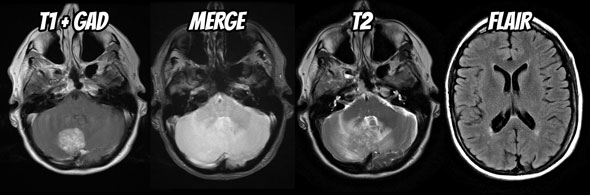
The trick is to first work out what the differential diagnosis for it is. Fortunately for this mass in a 60 year old female the likely differential is quite short: metastasis (common primaries include breast, lung, melanoma, RCC and GIT), hemangioblastoma and possibly a meningioma. We are not going to even consider an acoustic schwannoma or epidermoid as these are easily excluded by location and appearance.

Additionally as the patient is 60 years of age first presentation of von Hippel Lindau syndrome would be unusual, and it is probably safe to not dwell on it too much, lest you give the impression you do not know this.


Next you need to know which features are going to help you distinguish between them. They include:

  • large flow voids are common in hemangioblastomas
  • hemorrhage is common is RCC and melanoma metastases
  • broad dural base / dural tail are common in meningiomas (although can be seen in dural metastases e.g. breast cancer)
  • multiple lesions would favor metastases, or hemangioblastoma in the setting of von Hippel Lindau syndrome (unlikely in this age group).

You also need to think about management issues, the main one in this case is distortion of the fourth ventricle.


Relevant negatives in action

So here is how I would try and present his case. Relevant negatives are in bold.

  • Within the right cerebellar hemisphere is a rounded vividly enhancing mass with surrounding vasogenic edema. It exerts significant local mass effect, distorting the fourth ventricle but at this stage is not associated with obstructive hydrocephalus. The mass does not abut the dura, appearing intra-axial. It does not have prominent flow voids nor is there evidence of hemorrhage. No other similar lesions are seen either in the posterior fossa or elsewhere.

Then you can throw in some statements of normality if you really feel like it, although in this instance you have covered most things. You could go on and add:

  • The remainder of the scan is unremarkable for age.

Your next step would be your conclusion (or interpretation) and your reader / examiner won’t be at all surprised when you state:

  • Findings in this patient favor a solitary metastasis, and a lung or breast primary are most likely.

You don’t even need to go into why at this stage, because you have, by virtue of your description and judicious use of relevant negatives, already implied that you know the differential diagnosis and the important features of each.

It is important to note that sometimes a relevant negative does not preclude discussing a particular aspect of the case, and it would be prudent in this case to also add:

  • Despite no current obstructive hydrocephalus, urgent referral to a neurosurgeon is prudent and I would contact the treating physician to inform them of these findings.


Why is this so often done badly?

The problem with using relevant negatives well in the setting of an oral exam is that it requires you to have a differential very early on in the case, before you have described andy very much. This is usually the case by the time you get to your exam, but is usually not the case when you are starting out. So if have not been effectively practicing without wasting time or practicing your oral technique in the shower from the very start, you will not have been practicing the secret art of relevant negatives. The end result is that your descriptions will be longer, baggier, filled with seemingly random negatives and your examiner will be unsure of what you are going to say in your conclusion.

So, go back and practice. Look at your reports and the way you present cases and look for ways of introducing relevant negatives and removing irrelevant ones. This will have an enormous impact on how well you convey knowledge during an oral exam (see islands of knowledge vs puddles of ignorance) as well as allow you to create well crafted reports for the rest of your professional career.


Read next: Never surprise your examiner


Dr Frank Gaillard is a neuroradiologist at the Royal Melbourne Hospital, Melbourne, Australia, and is the Founder and Editor of Radiopaedia.org.

NB: Opinions expressed are those of the author alone, and are not those of his employer, or of Radiopaedia.org


Blog Subscription

We will only send you an email when there are new posts.

Updating… Please wait.

 Unable to process the form. Check for errors and try again.

 Thank you for updating your details.