O'Donoghue's unhappy triad
Diagnosis certain
Updates to Case Attributes
Body
was changed:
O'Donoghue described the injuries as:
- anterior cruciate ligament (ACL) tear
- medial collateral ligament (MCL) tear / sprain
- medial meniscal tear (lateral compartment bone bruise)
These images are from Dr. John Hunter's amazing MSK collection. Dr. John Hunter is a professor in the department of radiology (musculoskeletal section) atUC Davis School of Medicine.
This case was donated to Radiopaedia.org by Radswiki.net.
-<p>These images are from Dr. John Hunter's amazing MSK collection. Dr. John Hunter is a professor in the department of radiology (musculoskeletal section) at <a href="http://www.ucdmc.ucdavis.edu/radiology/">UC Davis School of Medicine</a>.</p><p>This case was donated to Radiopaedia.org by <a href="/radswikinet-1">Radswiki.net</a></p>- +<p>O'Donoghue described the injuries as:</p><ol>
- +<li><a href="/articles/anterior-cruciate-ligament-tear">anterior cruciate ligament (ACL) tear</a></li>
- +<li>
- +<a href="/articles/medial-collateral-ligament-of-the-knee">medial collateral ligament (MCL)</a> tear / sprain</li>
- +<li><a href="/articles/meniscal-tear">medial meniscal tear (lateral compartment bone bruise)</a></li>
- +</ol><p>These images are from Dr. John Hunter's amazing MSK collection. Dr. John Hunter is a professor in the department of radiology (musculoskeletal section) at <a href="http://www.ucdmc.ucdavis.edu/radiology/">UC Davis School of Medicine</a>.</p><p>This case was donated to Radiopaedia.org by <a href="/radswikinet-1">Radswiki.net</a>.</p>
Presentation
was added:
Twisting injury of the knee.
Updates to Study Attributes
Findings
was changed:
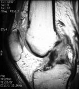
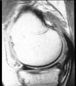
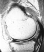
MR images demonstrate O'Donoghue's unhappy triad.
- wavy anterior cruciate ligament with interrupted proximal attachment, reflecting complete tear
- grade III tear of medial collateral ligament, with surrounding soft tissue oedema
- horizontal tear of the posterior horn of medial meniscus reaching the inferior articular surface
Mild knee effusion.
Bone marrow oedema of the lateral tibial plateau and lateral femoral condyle.
Images Changes:
Image MRI (PD) ( update )
Perspective
was set to
Sagittal.
Specifics
was set to
PD.
Image MRI (PD) ( update )
Perspective
was set to
Sagittal.
Specifics
was set to
PD.
Image MRI (PD) ( update )
Perspective
was set to
Sagittal.
Specifics
was set to
PD.
Image MRI (STIR) ( update )

Perspective
was set to
Coronal.
Specifics
was set to
STIR.