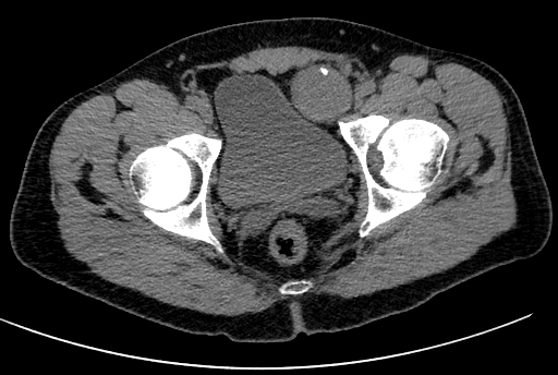
Non-enhanced CT images showed a high attenuation pedunculated solid mass in the left pelvis growing between the urinary bladder, left external iliac vessels and the left rectus abdominis muscle which is anteriorly displaced. The inguinal canal is tangentially encompassed by the mass which also compresses the external iliac vein against the iliopsoas muscle with a peduncle pointing toward it. Note the eccentric calcification in the antero-basal portion of the lesion where the mass abuts the postero-medial border of the inguinal canal close to the running course of the genitofemoral nerve where it probably comes from.