Extraskeletal chondrosarcoma
Diagnosis almost certain
Updates to Case Attributes
Status
changed from draft to published (public).
Published At
was set to
.
Body
was changed:
Case of biopsypathologically proven extraskeletal chondrosarcoma.
Case courtesy of Dr. George Matcuk.
-<p>Case of biopsy proven <a title="Extraskeletal chondrosarcoma" href="/articles/extraskeletal-chondrosarcoma-1">extraskeletal chondrosarcoma</a>. </p>- +<p>Case of pathologically proven <a href="/articles/extraskeletal-chondrosarcoma-1">extraskeletal chondrosarcoma</a>. </p><p> </p><p>Case courtesy of Dr. George Matcuk. </p>
Diagnostic Certainty
was set to
.
Updates to Study Attributes
Findings
was changed:
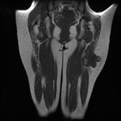
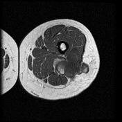
There is a large lobulated mass within the distal gluteus maximus near its insertion on the linea aspera, which invades the vastis lateralis and abuts the sciatic neurovascular bundle. This mass demonstrates predominately low T1 signal which is isointense to muscle and hyperintense on STIR, with scattered areas of fat signal. There is avid post-contrast enhancement of the lesion. No peri-tumoral edema is appreciated.
Images Changes:
Image MRI (T1) ( update )

Stack
was set to
.
Single Or Stack Root
was set to
.
Image MRI (T1) ( update )
Stack
was set to
.
Single Or Stack Root
was set to
.
Image MRI (T1) ( update )

Stack
was set to
.
Single Or Stack Root
was set to
.
Image MRI (T1) ( update )
Stack
was set to
.
Single Or Stack Root
was set to
.
Image MRI (STIR) ( update )

Stack
was set to
.
Single Or Stack Root
was set to
.
Image MRI (STIR) ( update )

Stack
was set to
.
Single Or Stack Root
was set to
.