Cardiac hemangioma
Diagnosis certain
Updates to Case Attributes
Age
changed from 60-65Y to 65 years.
Body
was changed:
Cardiac hemangioma is is an uncommon benign cardiac tumor tumour (5-10%). They have no typical age of presentation and no typical cardiac chamber predilection.
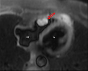
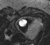
The appearance, in this case, is typical with hyperintense T2 signal and gradual postcontrast enhancement on the perfusion sequence.
-<p>Cardiac hemangioma is an uncommon benign cardiac tumor (5-10%). They have no typical age of presentation and no typical cardiac chamber predilection.</p><p>The appearance in this case is typical with hyperintense T2 signal and gradual postcontrast enhancement on the perfusion sequence.</p>- +<p>Cardiac hemangioma is an uncommon benign cardiac tumour (5-10%). They have no typical age of presentation and no typical cardiac chamber predilection.</p><p>The appearance, in this case, is typical with hyperintense T2 signal and gradual postcontrast enhancement on the perfusion sequence.</p>
Updates to Study Attributes
Findings
was changed:
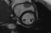
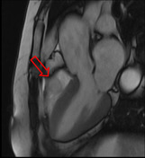
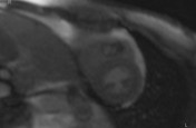
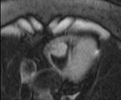
MRI images of a biopsy-proven cardiac cardiac hemangioma.
Intraluminal mass arising from the RV free wall, hyperintense in T2 weighted images with classic hemangioma-like discontinuous nodular enhancement.
Images Changes:
Image MRI (Cine SSFP) ( update )
Description
was removed:
Image MRI (Cine SSFP) ( update )
Description
was removed:
Image MRI (HASTE dark blood fat sat) ( update )
Description
was removed:
Image MRI (T2 dark blood fat sat) ( update )
Description
was removed:
Image MRI (postcontrast perfusion SSFP) ( update )
Description
was removed:
Image MRI (postcontrast T1 dark blood fat sat) ( update )
Description
was removed: