Cellular angiofibroma - vulva (histology)
Updates to Case Attributes
Cellular angiofibroma is a benign soft tissue lesion found in the inguinal, vulval or scrotal regions, usually in middle aged+ adults. The cells are myofibroblastic in nature. They are well circumscribed-circumscribed and comprise spindle cells with morphology described above, and show variable mitotic activity (not atypical, and most often minimal in number). Scattered mast cells are often also present and there is a fine fibrocollagenous stroma with no necrosis. They usually show small prominent vessels with hyalinised walls, which this case doesn't show well, however, the morphology and immunohistochemical findings fit with this diagnosis rather than one of the other differential diagnoses possible in this location:
-
Angiomyofibroblastoma: Usually also well
circumscribed-circumscribed, variable cellularity - paucicellular background with clustering of rounded cells around smaller, thin-walled vessels, desmin+ -
Aggressive angiomyxoma: Infiltrative, aggressively growing lesion, much less cellular with a more myxoid
/ loose/loose appearing background, desmin+ -
Leiomyoma:
FasicularFascicular growth pattern, with cells containing more cytoplasm and nuclear features more consistent with smooth muscle cells (plumper, blunt-ended cigarette nuclei), desmin+ - Solitary Fibrous Tumour: Same spindly cells, but hypo- and hyper-cellular areas, hyalinised stroma between cells, CD34+
These lesions are usually benign and do not recur, however, case reports of malignant forms have been published.
-<p>Cellular angiofibroma is a benign soft tissue lesion found in the inguinal, vulval or scrotal regions, usually in middle aged+ adults. The cells are myofibroblastic in nature. They are well circumscribed and comprise spindle cells with morphology described above, and show variable mitotic activity (not atypical, and most often minimal in number). Scattered mast cells are often also present and there is a fine fibrocollagenous stroma with no necrosis. They usually show small prominent vessels with hyalinised walls, which this case doesn't show well, however, the morphology and immunohistochemical findings fit with this diagnosis rather than one of the other <strong>differential diagnoses possible in this location</strong>:</p><ul>- +<p>Cellular angiofibroma is a benign soft tissue lesion found in the inguinal, vulval or scrotal regions, usually in middle aged+ adults. The cells are myofibroblastic in nature. They are well-circumscribed and comprise spindle cells with morphology described above, and show variable mitotic activity (not atypical, and most often minimal in number). Scattered mast cells are often also present and there is a fine fibrocollagenous stroma with no necrosis. They usually show small prominent vessels with hyalinised walls, which this case doesn't show well, however, the morphology and immunohistochemical findings fit with this diagnosis rather than one of the other <strong>differential diagnoses possible in this location</strong>:</p><ul>
-<strong>Angiomyofibroblastoma:</strong> Usually also well circumscribed, variable cellularity - paucicellular background with clustering of rounded cells around smaller, thin-walled vessels, desmin+</li>- +<strong><a title="Angiomyofibroblastoma" href="/articles/angiomyofibroblastoma">Angiomyofibroblastoma</a>:</strong> Usually also well-circumscribed, variable cellularity - paucicellular background with clustering of rounded cells around smaller, thin-walled vessels, desmin+</li>
-<strong><a title="Aggressive angiomyxoma" href="/articles/aggressive-angiomyxoma">Aggressive angiomyxoma</a>:</strong> Infiltrative, aggressively growing lesion, much less cellular with a more myxoid / loose appearing background, desmin+</li>- +<strong><a href="/articles/aggressive-angiomyxoma">Aggressive angiomyxoma</a>:</strong> Infiltrative, aggressively growing lesion, much less cellular with a more myxoid/loose appearing background, desmin+</li>
-<strong><a title="Uterine leiomyoma" href="/articles/uterine-leiomyoma">Leiomyoma</a>:</strong> Fasicular growth pattern, with cells containing more cytoplasm and nuclear features more consistent with smooth muscle cells (plumper, blunt-ended cigarette nuclei), desmin+</li>- +<strong><a href="/articles/uterine-leiomyoma">Leiomyoma</a>:</strong> Fascicular growth pattern, with cells containing more cytoplasm and nuclear features more consistent with smooth muscle cells (plumper, blunt-ended cigarette nuclei), desmin+</li>
-<strong><a title="Solitary fibrous tumour" href="/articles/solitary-fibrous-tumour">Solitary Fibrous Tumour</a>:</strong> Same spindly cells, but hypo- and hyper-cellular areas, hyalinised stroma between cells, CD34+</li>- +<strong><a href="/articles/solitary-fibrous-tumour">Solitary Fibrous Tumour</a>:</strong> Same spindly cells, but hypo- and hyper-cellular areas, hyalinised stroma between cells, CD34+</li>
Updates to Study Attributes
Image Pathology (H&E) ( update )
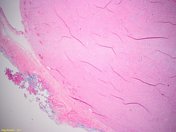
Image Pathology (H&E) ( update )

Image Pathology (H&E) ( update )

Image Pathology (H&E) ( update )

Image Pathology (H&E) ( update )

Image Pathology (C-kit) ( update )

Image Pathology (ER) ( update )
