Caput medusa
Updates to Case Attributes
Caput medusa (head of Medusa with multiple serpent-like veins in the umbilicus) reflects portal hypertension with the umbilicus being the end point for the recanalized paraumbilical vein (in this case highlighted by the fat containing-containing umbilical hernia). These veins are at higher pressure than normal thus are prone to haemorrhage with minimal trauma. Indeed in some cases, this can be fatal due to the associated coagulopathy of chronic liver disease.
-<p>Caput medusa (head of Medusa with multiple serpent-like veins in the umbilicus) reflects portal hypertension with the umbilicus being the end point for the recanalized paraumbilical vein (in this case highlighted by the fat containing umbilical hernia). These veins are at higher pressure than normal thus are prone to haemorrhage with minimal trauma. Indeed in some cases this can be fatal due to the associated coagulopathy of chronic liver disease.</p>- +<p>Caput medusa (head of Medusa with multiple serpent-like veins in the umbilicus) reflects portal hypertension with the umbilicus being the end point for the recanalized paraumbilical vein (in this case highlighted by the fat-containing umbilical hernia). These veins are at higher pressure than normal thus are prone to haemorrhage with minimal trauma. Indeed in some cases, this can be fatal due to the associated coagulopathy of chronic liver disease.</p>
References changed:
- 1. Bott E, O'Donnell C, Burke M. Post mortem CT demonstration of hemoperitoneum caused by rupture of a paraumbilical vein into a paraumbilical hernia in a man with liver cirrhosis and portal hypertension. Forensic science, medicine, and pathology. 9 (1): 77-81. <a href="https://doi.org/10.1007/s12024-012-9384-4">doi:10.1007/s12024-012-9384-4</a> - <a href="https://www.ncbi.nlm.nih.gov/pubmed/23055059">Pubmed</a> <span class="ref_v4"></span>
- Bott E, O'Donnell C, Burke M. Post mortem CT demonstration of hemoperitoneum caused by rupture of a paraumbilical vein into a paraumbilical hernia in a man with liver cirrhosis and portal hypertension. Forensic science, medicine, and pathology. 9 (1): 77-81. <a href="https://doi.org/10.1007/s12024-012-9384-4">doi:10.1007/s12024-012-9384-4</a> - <a href="https://www.ncbi.nlm.nih.gov/pubmed/23055059">Pubmed</a> <span class="ref_v4"></span>
Updates to Study Attributes
Caput medusa (i.e. leash of abnormal veins).
Image Annotated image (Sagittal) ( update )
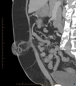
Image Annotated image (Axial) ( update )

Image Annotated image (Medusa by Caravaggio) ( update )

Image Annotated image (thick MIP coronal) ( update )
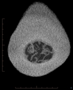
Image 1 Annotated image (Axial) ( update )
Image 2 Annotated image (Sagittal) ( update )
Image 3 Annotated image (thick MIP coronal) ( update )
Updates to Study Attributes
Features of cirrhosis and portal hypertension with multiple venous collaterals especially around the spleen (porto-systemic(portosystemic shunts). Recannalized paraumbilical vein entering an umbilical hernia containing fat where there is a caput medusa (i.e. leash of abnormal veins)
Image CT (C+ portal venous phase) ( update )

Image CT (C+ portal venous phase) ( update )
