Retained gallstone
Diagnosis certain
Updates to Case Attributes
Age
changed from 48 to 50 years.
Body
was changed:
PatientThe patient was status post cholecystectomy about one year ago and had continued right abdominal pain. Multiple follow up abdominal CTs were performed in which there was no mention of the retained gallstones. Eventually, an abdominal MRI was performed in which a presumed diagnosis of retained gallstones was made. This was confirmed by surgical exploration and removal of the stones.
-<p>Patient was status post <a title="Cholecystectomy" href="/articles/cholecystectomy-1">cholecystectomy</a> about one year ago and had continued right abdominal pain. Multiple follow up abdominal CTs were performed in which there was no mention of the <a title="Retained gallstones" href="/articles/retained-gallstone">retained gallstones</a>. Eventually, an abdominal MRI was performed in which a presumed diagnosis of retained gallstones was made. This was confirmed by surgical exploration and removal of the stones.</p>- +<p>The patient was status post <a href="/articles/cholecystectomy-1">cholecystectomy</a> about one year ago and had continued right abdominal pain. Multiple follow up abdominal CTs were performed in which there was no mention of the <a href="/articles/retained-gallstone">retained gallstones</a>. Eventually, an abdominal MRI was performed in which a presumed diagnosis of retained gallstones was made. This was confirmed by surgical exploration and removal of the stones.</p>
Updates to Study Attributes
Findings
was changed:
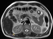
There are a couple of adjacent focal T1 hyperintensities in the hepatorenal recess, with corresponding T2 hypointensity (yellow arrows), representing retained (dropped) gallstones, which was confirmed by surgery.
Images Changes:
Image Annotated image (Axial T1 fat sat annotated ) ( update )

Description
was removed:
Image Annotated image (Axial T2 annotated ) ( update )
Description
was removed: